Copyright
©2014 Baishideng Publishing Group Co.
World J Gastroenterol. Apr 21, 2014; 20(15): 4335-4340
Published online Apr 21, 2014. doi: 10.3748/wjg.v20.i15.4335
Published online Apr 21, 2014. doi: 10.3748/wjg.v20.i15.4335
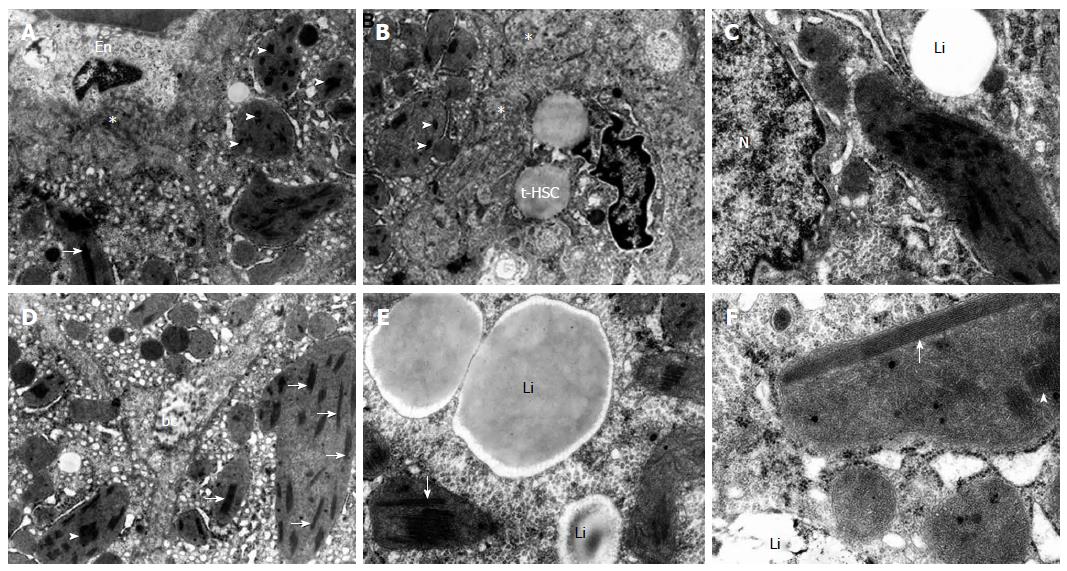
Figure 1 Ultrastructural appearance of polymorphic hepatic mitochondria, with megamitochondria at the foreground, containing linear crystalline inclusions and reduced cristae in bioptates from different pediatric patients with non-alcoholic steatohepatitis.
Abnormal mitochondria distributed randomly in topographically varied parts of hepatocytes. A-F: Linear inclusions - cut longitudinally (arrows) and in cross section (arrowheads) in the matrix of moderately or markedly increased electron density in the majority of altered mitochondria, especially elongated and rounded megamitochondria. A, B: Megamitochondria (MMC) located in the vascular pole of hepatocytes; the mitochondrial lesions are accompanied by the proliferation of smooth endoplasmic reticulum; considerably swollen endothelial cell (En) of the sinusoidal vessel; transitional hepatic stellate cells (t-HSC), accumulation of electron-dense material, and collagen formed (asterisk) in the dilated space of Disse (original magnification × 7000); C: The MMC located in the nuclear region of hepatocyte is accompanied by glycogen accumulation; the megamitochondrium shows enlarged intramitochondrial dense granules in the vicinity of linear crystalline inclusions; hepatocyte nucleus (N); lipid material (Li) within hepatocyte cytoplasm (original magnification × 12000); D: MMC distributed randomly within the biliary pole of hepatocytes accompanied by the proliferation of smooth endoplasmic reticulum; bc - biliary canaliculus (original magnification × 7000); E, F: MMC located at a certain distance from the nucleus of hepatocyte; mitochondrial abnormalities are accompanied by glycogen accumulation; some MMC show enlarged intramitochondrial dense granules in the vicinity of linear crystalline inclusions; lipid material (Li) (original magnification × 12000, × 20000, respectively).
- Citation: Lotowska JM, Sobaniec-Lotowska ME, Bockowska SB, Lebensztejn DM. Pediatric non-alcoholic steatohepatitis: The first report on the ultrastructure of hepatocyte mitochondria. World J Gastroenterol 2014; 20(15): 4335-4340
- URL: https://www.wjgnet.com/1007-9327/full/v20/i15/4335.htm
- DOI: https://dx.doi.org/10.3748/wjg.v20.i15.4335


