Copyright
©2014 Baishideng Publishing Group Co.
World J Gastroenterol. Mar 21, 2014; 20(11): 2810-2824
Published online Mar 21, 2014. doi: 10.3748/wjg.v20.i11.2810
Published online Mar 21, 2014. doi: 10.3748/wjg.v20.i11.2810
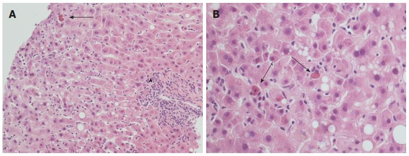
Figure 1 Typical histopathological appearance of acute recurrent hepatitis C.
A: Lobular architectural disarray, lobular necrosis with lymphocytic sinusoidal infiltrate and visible Councilman bodies (black arrow) are evident, as well as a mild portal tract inflammation (arrowhead); hematoxylin-eosin stain, × 20 magnification; B: Detail of the same case at × 40 magnification: note the high number of Councilman bodies in a single field (black arrows), and a minimal amount of macrovesicular steatosis.
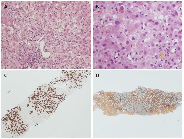
Figure 2 Histopathological appearance of fibrosing cholestatic recurrent hepatitis C.
Hematoxylin-eosin stain × 10 (A) and × 40 (B) magnification: note the lobular architectural disarray, the portal tract fibrosis and distortion, the lobular necrosis with Councilman bodies and the cholestasis with hepatocellular feathery degeneration and ballooning. The immunohistochemistry for keratin 19 (C) highlights the prominent ductular reaction, while the reticulin stain (D) indicates advanced fibrosis.
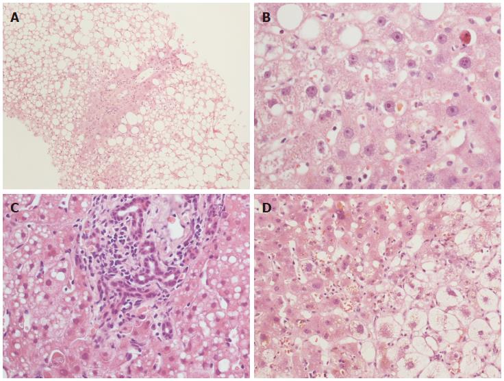
Figure 3 Histopathological features with a prognostic significance in recurrent hepatitis C.
Steatosis (A) and cholestasis (B) are well shown. Cholestasis can be associated with bile duct proliferation (C) and/or hepatocellular ballooning (D). Hematoxylin-eosin stain, × 10 (A) and × 40 (B-D) magnification.
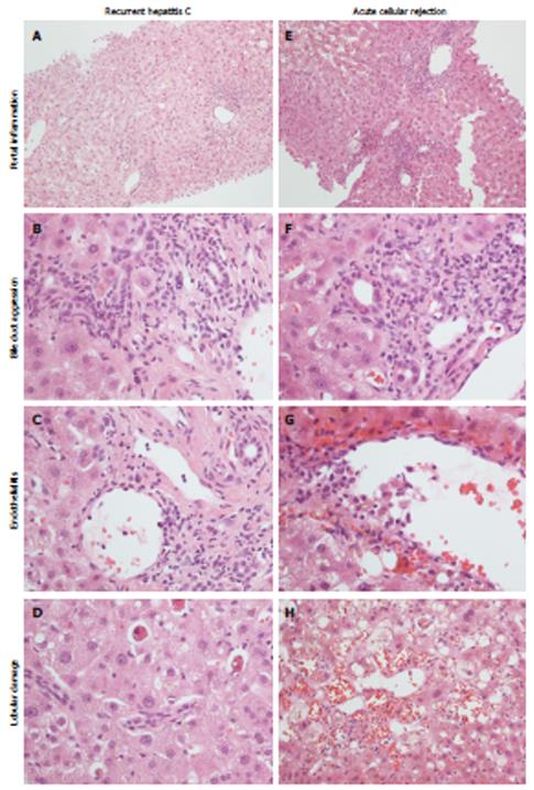
Figure 4 Histopathological appearance of recurrent hepatitis C (left) and acute cellular rejection (right).
Portal inflammation is commonly mild in recurrent hepatitis C (A), with a predominant lympho-monocytic infiltrate and mild bile duct invasion (B), while in acute cellular rejection there is a mixed and more pronounced inflammatory infiltrate (E), with evident bile duct invasion (F). Endothelialitis can be found in both conditions (C, G). Lobular necrosis is more typical of recurrent hepatitis C (D); in acute cellular rejection hemorrhage and sinusoid dilatation are more evident (H).
- Citation: Vasuri F, Malvi D, Gruppioni E, Grigioni WF, D’Errico-Grigioni A. Histopathological evaluation of recurrent hepatitis C after liver transplantation: A review. World J Gastroenterol 2014; 20(11): 2810-2824
- URL: https://www.wjgnet.com/1007-9327/full/v20/i11/2810.htm
- DOI: https://dx.doi.org/10.3748/wjg.v20.i11.2810


