Copyright
©The Author(s) 1996.
World J Gastroenterol. Dec 15, 1996; 2(4): 218-219
Published online Dec 15, 1996. doi: 10.3748/wjg.v2.i4.218
Published online Dec 15, 1996. doi: 10.3748/wjg.v2.i4.218
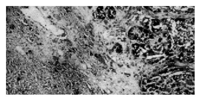
Figure 1 Staining intensity in hepatocellular carcinoma tissue (right panel) is higher than that in the adjacent nontumorous liver tissue (left panel).
(Magnification, × 100)
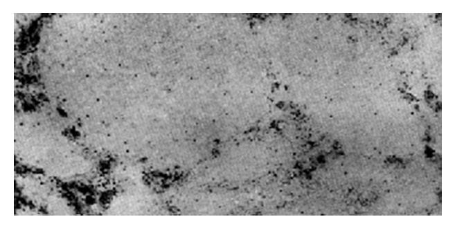
Figure 2 Cancerous nodules showing negative staining.
(Magnification, × 100)
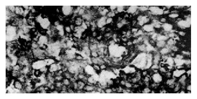
Figure 3 The positive signal of nm23-H1 mRNA is mainly distributed in the cytoplasm.
(Magnification, × 200)
- Citation: Chen XD, Dai YM, Yang JM, Bao JZ, Wang JJ, Chong WM. Expression of metastasis suppressor gene nm23 in human hepatocellular carcinoma. World J Gastroenterol 1996; 2(4): 218-219
- URL: https://www.wjgnet.com/1007-9327/full/v2/i4/218.htm
- DOI: https://dx.doi.org/10.3748/wjg.v2.i4.218


