Copyright
©The Author(s) 1996.
World J Gastroenterol. Mar 25, 1996; 2(1): 30-33
Published online Mar 25, 1996. doi: 10.3748/wjg.v2.i1.30
Published online Mar 25, 1996. doi: 10.3748/wjg.v2.i1.30
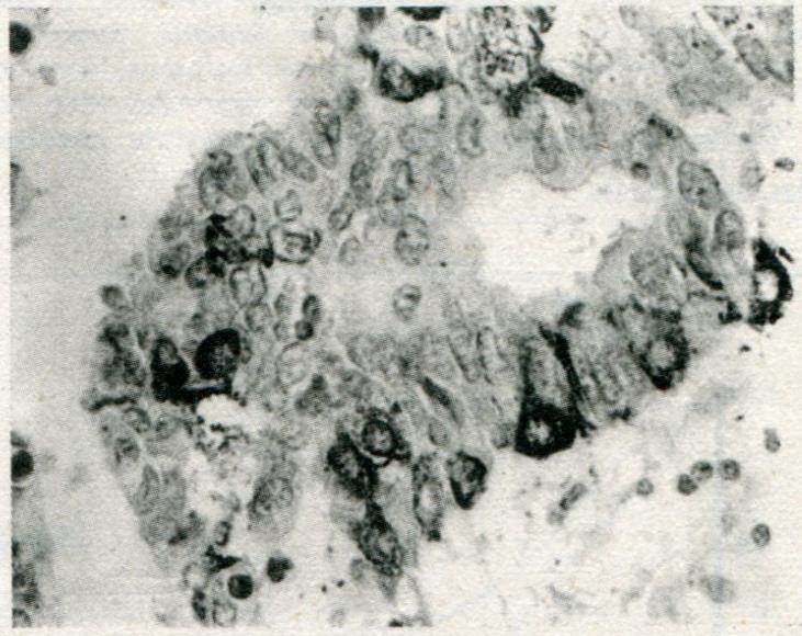
Figure 1 Triangular, flat, or cylindrical neuroendocrine cells with Chromogranin A positivity at the infranuclear spaces.
ABC method, magnification × 400.
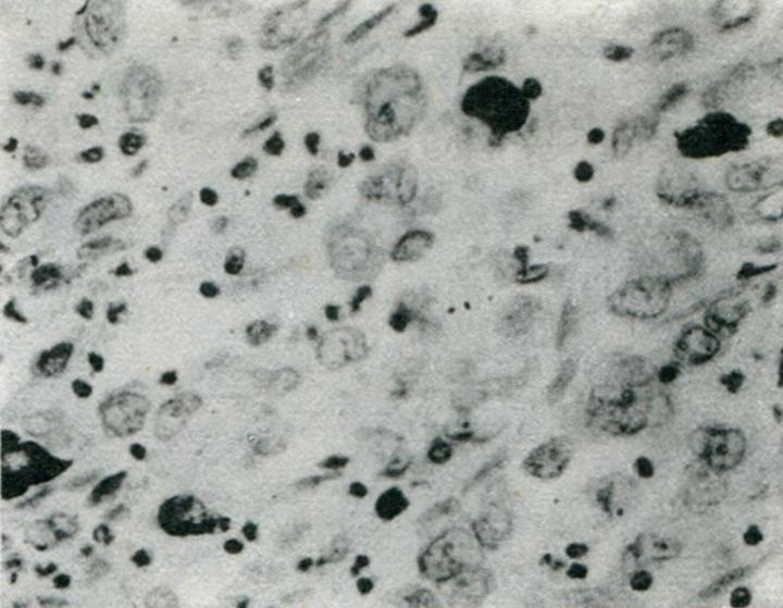
Figure 2 HCG-positive cells in poorly differentiated gastric carcinoma.
ABC method, magnification × 400.
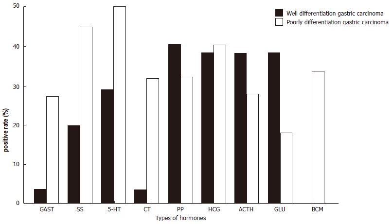
Figure 3 Hormone containing neuroendocrine cells in gastric carcinoma and cancer differentiation.
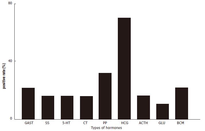
Figure 4 Hormone-positive neuroendocrine cells in gastric carcinoma with lymph nodes metastasis.
Figure 5 Survival curve of 54 gastric carcinoma patients.
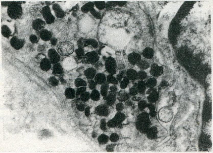
Figure 6 Type I neuroendocrine cell with 100-200 nm-sized pleomorphic granules.
Transmission electron microscope, magnification × 20000.
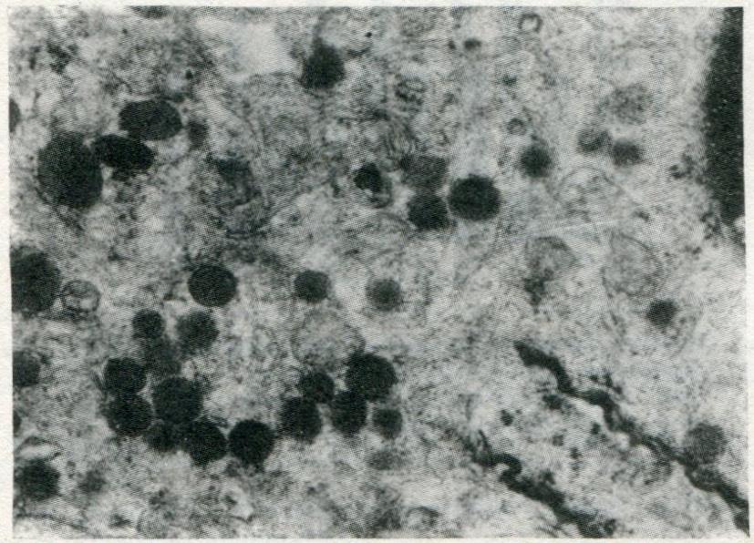
Figure 7 neuroendocrine cell showing round granular dense core surrounded by a membrane.
Transmission electron microscope, magnification × 20000.
- Citation: Wang LP, Yu JY, Shi JQ, Liang YJ. Clinico-pathologic significance of neuroendocrine cells in gastric cancer tissue. World J Gastroenterol 1996; 2(1): 30-33
- URL: https://www.wjgnet.com/1007-9327/full/v2/i1/30.htm
- DOI: https://dx.doi.org/10.3748/wjg.v2.i1.30


