Copyright
©2013 Baishideng Publishing Group Co.
World J Gastroenterol. Feb 28, 2013; 19(8): 1314-1317
Published online Feb 28, 2013. doi: 10.3748/wjg.v19.i8.1314
Published online Feb 28, 2013. doi: 10.3748/wjg.v19.i8.1314
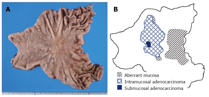
Figure 1 Macroscopic finding and distribution of ectopic cystic lesion and cancer in surgically excised stomach.
Surgically excised stomach (A) and schematic distribution of the lesions (B). Ectopic cystic mucosa is located mainly on the oral side and partly in the body part overlapped with a cancer lesion (shaded area). A well- to moderately-differentiated tubular adenocarcinoma in the intramucosal layer (checked area) and part of a poorly-differentiated component infiltrating the submucosal layer (blue area) are located mainly in the body part of the stomach.
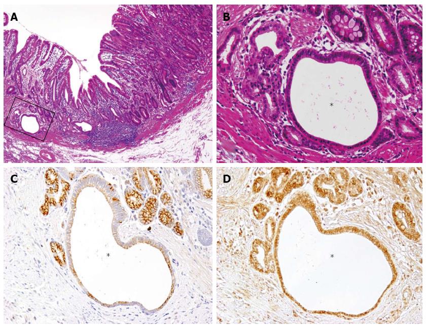
Figure 2 Expression of KCNE2 and estrogen receptor in non-neoplastic cystic lesion by immunohistochemistry.
A: Low power magnification of a transitional area from the non-neoplastic mucosal layer with intramucosal cystic lesions (left) to the intramucosal adenocarcinoma (right, HE, × 40); B: High power magnification (A, squared area) around the intramucosal cystic lesion (asterisk, HE, × 200); C: KCNE2 immunostaining of a serial section of (B) shows that KCNE2 is almost negative in the dilated cystic gland (asterisk), while the surrounding non-cystic glands are positive (× 200); D: Estrogen receptor immunostaining of serial sections of (B) and (C) show that ER is equally expressed in both cystic (asterisk) and non-cystic glands (× 200).
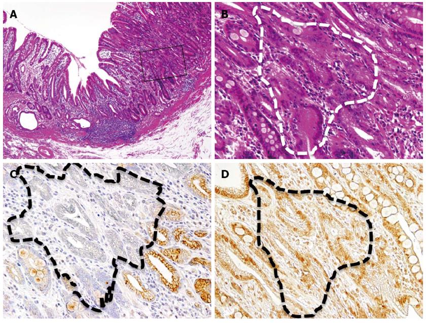
Figure 3 Expression of KCNE2 and estrogen receptor in adenocarcinoma area.
A: Low power magnification of a transitional area from the non-neoplastic mucosal layer with intramucosal cystic lesions (left) to the intramucosal adenocarcinoma (right, HE, × 40); B: High power magnification (A, squared area) around the intramucosal adenocarcinoma (circled by white-hatched line, HE, × 200); C: KCNE2 immunostaining of a serial section of (B) shows that KCNE2 expression is almost negative in adenocarcinoma (circled by black-hatched line), while the surrounding non-neoplastic glands are positive (× 200); D: Estrogen receptor immunostaining of serial sections of (B) and (C) show that estrogen receptor is equally expressed in both cancerous (circled by black-hatched line) and non-neoplastic glands (× 200).
- Citation: Kuwahara N, Kitazawa R, Fujiishi K, Nagai Y, Haraguchi R, Kitazawa S. Gastric adenocarcinoma arising in gastritis cystica profunda presenting with selective loss of KCNE2 expression. World J Gastroenterol 2013; 19(8): 1314-1317
- URL: https://www.wjgnet.com/1007-9327/full/v19/i8/1314.htm
- DOI: https://dx.doi.org/10.3748/wjg.v19.i8.1314


