Copyright
©2013 Baishideng Publishing Group Co.
World J Gastroenterol. Feb 7, 2013; 19(5): 778-780
Published online Feb 7, 2013. doi: 10.3748/wjg.v19.i5.778
Published online Feb 7, 2013. doi: 10.3748/wjg.v19.i5.778
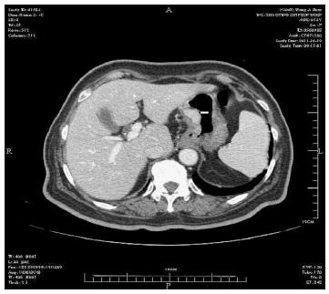
Figure 1 Enhanced image of nodule located in the lesser gastric curvature.
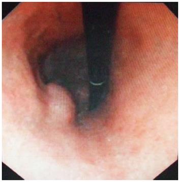
Figure 2 Elevated nodule on the lesser curvature side of the gastric antrum.
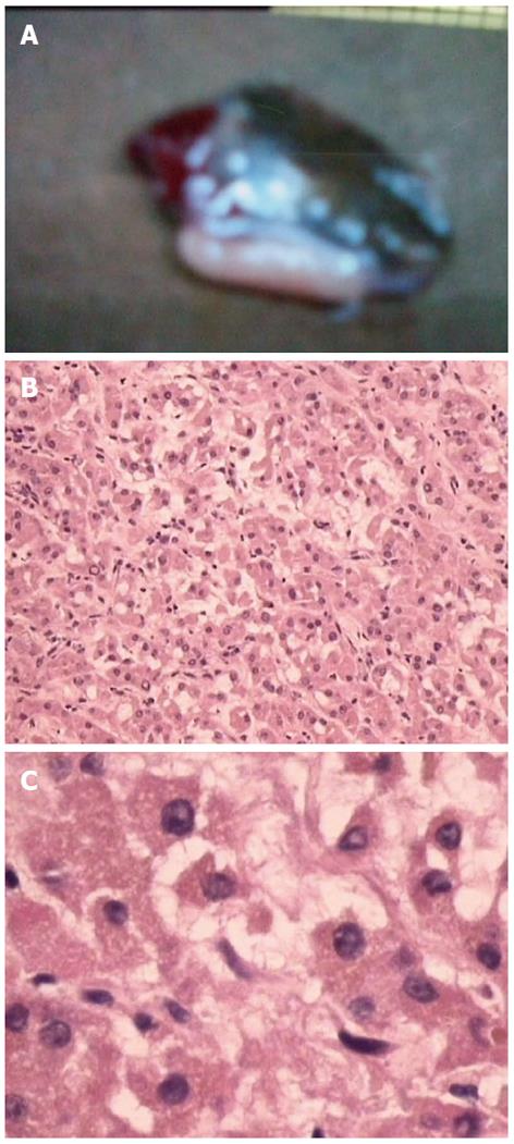
Figure 3 Analysis of tumor cells.
A: Gastric wall mass, approximately 20 mm × 30 mm in size; B: Tumor cells under low power magnification; C: Tumor cells under high power magnification.
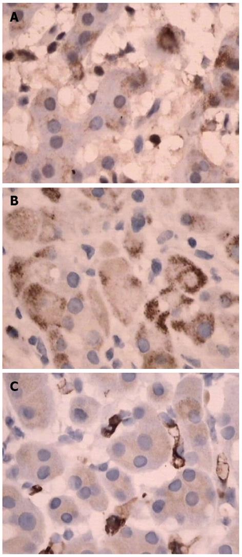
Figure 4 Immunohistochemistry.
A: S-100 basement cell negative; B: Melan-A positive; C: CD34 positive.
- Citation: Ren PT, Fu H, He XW. Ectopic adrenal cortical adenoma in the gastric wall: Case report. World J Gastroenterol 2013; 19(5): 778-780
- URL: https://www.wjgnet.com/1007-9327/full/v19/i5/778.htm
- DOI: https://dx.doi.org/10.3748/wjg.v19.i5.778


