Copyright
©2013 Baishideng Publishing Group Co.
World J Gastroenterol. Dec 14, 2013; 19(46): 8789-8792
Published online Dec 14, 2013. doi: 10.3748/wjg.v19.i46.8789
Published online Dec 14, 2013. doi: 10.3748/wjg.v19.i46.8789
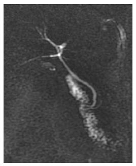
Figure 1 Magnetic resonance of abdomen and bile duct-nuclear magnetic resonance.
Images of bile duct-nuclear magnetic resonance demonstrating the biliary tree without evidence of obstructive processes.
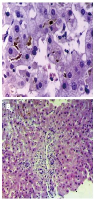
Figure 2 Transcutaneous liver biopsy guided by ultrasound.
A: Liver biopsy at x 100 magnification - moderate cholestasis is demonstrated associated with discrete parenchymal activity with slight tumefaction of hepatocytes; B: Liver biopsy at × 40 magnification - portal space is shown with discrete increase in periportal lymphocytes, extravasation of lymphocytes towards the interface (spillover), absence of piecemeal necrosis and cholestasis.
- Citation: Beraldo DO, Melo JF, Bonfim AV, Teixeira AA, Teixeira RA, Duarte AL. Acute cholestatic hepatitis caused by amoxicillin/clavulanate. World J Gastroenterol 2013; 19(46): 8789-8792
- URL: https://www.wjgnet.com/1007-9327/full/v19/i46/8789.htm
- DOI: https://dx.doi.org/10.3748/wjg.v19.i46.8789


