Copyright
©2013 Baishideng Publishing Group Co.
World J Gastroenterol. Dec 7, 2013; 19(45): 8445-8448
Published online Dec 7, 2013. doi: 10.3748/wjg.v19.i45.8445
Published online Dec 7, 2013. doi: 10.3748/wjg.v19.i45.8445
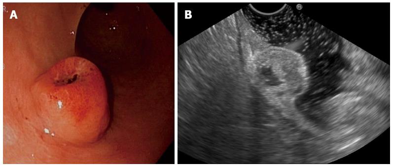
Figure 1 Endoscopic view.
A: Showing a subepithelial lesion with ulceration and blood clotting; B: Showing a hypoechoic lesion arising from the second and third layers of the gastric wall, with a central anechoic area.
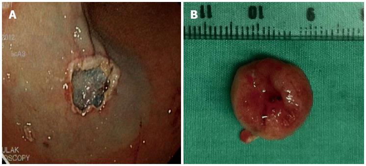
Figure 2 Lesion was removed in an en-bloc fashion by an electrical snare.
A: Submucosal layer after completion of the endoscopic mucosal resection; B: En-bloc resected tissue.
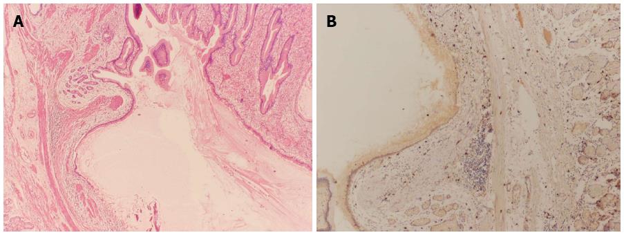
Figure 3 Histopathology and immunohistochemistry showing.
A: The eroded gastric mass composed of dilated gastric glands surrounded by fibromuscular tissue, fibroblasts, and smooth muscle bundles; B: CD117-negative staining.
- Citation: Deesomsak M, Aswakul P, Junyangdikul P, Prachayakul V. Rare adult gastric duplication cyst mimicking a gastrointestinal stromal tumor. World J Gastroenterol 2013; 19(45): 8445-8448
- URL: https://www.wjgnet.com/1007-9327/full/v19/i45/8445.htm
- DOI: https://dx.doi.org/10.3748/wjg.v19.i45.8445


