Copyright
©2013 Baishideng Publishing Group Co.
World J Gastroenterol. Nov 21, 2013; 19(43): 7820-7824
Published online Nov 21, 2013. doi: 10.3748/wjg.v19.i43.7820
Published online Nov 21, 2013. doi: 10.3748/wjg.v19.i43.7820
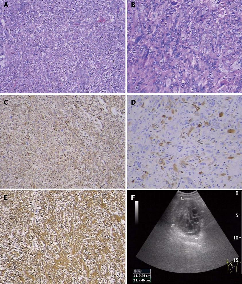
Figure 1 Hematoxylin and eosin stained sections, immunohistochemical test and ultrasonography diagnosis.
A: Histologic findings of the tumor; the morphology of sarcomatoid carcinoma of the pancreas (SCP) is shown (hematoxylin and eosin, × 100); B: Microscopically, the excised tumor tissue comprised cancer cells and mesenchymal cells, with dispersion of atypical cells and obvious karyokinesis (hematoxylin and eosin, × 200); C: Widely diffuse immunohistochemical staining for the epithelial marker cytokeratin 18 (× 100); D: Heterogeneous immunohistochemical staining for the epithelial marker pan-cytokeratin (Pan-CK) (× 100); E: Widely diffuse immunohistochemical staining for the mesenchymal marker vimentin (× 100); F: Ultrasonography revealed a 93 mm × 94 mm × 75 mm mass of mixed echogenicity in the tail of the pancreas.
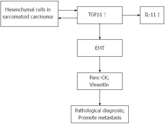
Figure 2 Mechanism of transforming growth factorβ1 regulating the epithelial-to-mesenchymal transition of sarcomatoid carcinoma of the pancreas.
TGF: Transforming growth factor; EMT: Epithelial-to-mesenchymal transition; IL-11: Interleukin-11.
- Citation: Ren CL, Jin P, Han CX, Xiao Q, Wang DR, Shi L, Wang DX, Chen H. Unusual early-stage pancreatic sarcomatoid carcinoma. World J Gastroenterol 2013; 19(43): 7820-7824
- URL: https://www.wjgnet.com/1007-9327/full/v19/i43/7820.htm
- DOI: https://dx.doi.org/10.3748/wjg.v19.i43.7820


