Copyright
©2013 Baishideng Publishing Group Co.
World J Gastroenterol. Nov 7, 2013; 19(41): 7205-7208
Published online Nov 7, 2013. doi: 10.3748/wjg.v19.i41.7205
Published online Nov 7, 2013. doi: 10.3748/wjg.v19.i41.7205
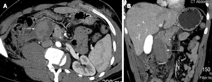
Figure 1 Axial image (A) and coronal image (B) from abdominal computed tomography with contrast depicts the intramural hematoma as it compresses the head of the pancreas and its’ associated hemorrhage as it extends into the retroperitoneum (arrows).
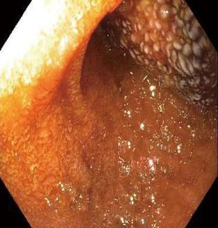
Figure 2 Esophagogastroduodenoscopy showing near complete obstruction of duodenum by intramural hematoma.
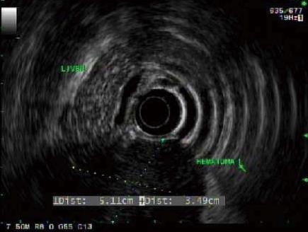
Figure 3 Endoscopic ultrasound image of intramural duodenal hematoma.
- Citation: Eichele DD, Ross M, Tang P, Hutchins GF, Mailliard M. Spontaneous intramural duodenal hematoma in type 2B von Willebrand disease. World J Gastroenterol 2013; 19(41): 7205-7208
- URL: https://www.wjgnet.com/1007-9327/full/v19/i41/7205.htm
- DOI: https://dx.doi.org/10.3748/wjg.v19.i41.7205


