Copyright
©2013 Baishideng Publishing Group Co.
World J Gastroenterol. Oct 28, 2013; 19(40): 6943-6946
Published online Oct 28, 2013. doi: 10.3748/wjg.v19.i40.6943
Published online Oct 28, 2013. doi: 10.3748/wjg.v19.i40.6943
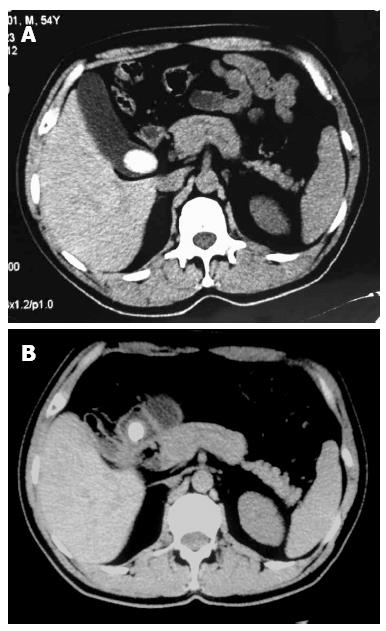
Figure 1 Abdominal computed tomography.
A: Revealing a large calcified mass in the gallbladder (one year prior); B: Indicating the migration of the large stone from the gallbladder to the pylorus and gas shadows seen in the gallbladder (this time).
- Citation: Yang D, Wang Z, Duan ZJ, Jin S. Laparoscopic treatment of an upper gastrointestinal obstruction due to Bouveret’s syndrome. World J Gastroenterol 2013; 19(40): 6943-6946
- URL: https://www.wjgnet.com/1007-9327/full/v19/i40/6943.htm
- DOI: https://dx.doi.org/10.3748/wjg.v19.i40.6943


