Copyright
©2013 Baishideng Publishing Group Co.
World J Gastroenterol. Oct 28, 2013; 19(40): 6842-6848
Published online Oct 28, 2013. doi: 10.3748/wjg.v19.i40.6842
Published online Oct 28, 2013. doi: 10.3748/wjg.v19.i40.6842
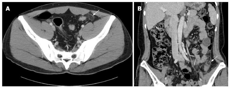
Figure 1 Computer tomography image of a 31-year-old man who presented with acute left lower quadrant pain.
A: A 31-year-old man who presented with acute left lower quadrant pain. An oval fatty mass with a hyperattenuated ring and surrounding inflammation adjacent to the sigmoid colon (arrow) is noted. The lesion corresponds to the site of tenderness and is characteristic of primary epiploic appendagitis. B: A 48-year-old female who presented with left lower quadrant pain. An ovoid fat attenuated mass with a central high attenuation area within the inflamed epiploic appendage in the distal descending colon (arrow) is shown.
-
Citation: Hwang JA, Kim SM, Song HJ, Lee YM, Moon KM, Moon CG, Koo HS, Song KH, Kim YS, Lee TH, Huh KC, Choi YW, Kang YW, Chung WS. Differential diagnosis of left-sided abdominal pain: Primary epiploic appendagitis
vs colonic diverticulitis. World J Gastroenterol 2013; 19(40): 6842-6848 - URL: https://www.wjgnet.com/1007-9327/full/v19/i40/6842.htm
- DOI: https://dx.doi.org/10.3748/wjg.v19.i40.6842


