Copyright
©2013 Baishideng Publishing Group Co.
World J Gastroenterol. Oct 28, 2013; 19(40): 6794-6804
Published online Oct 28, 2013. doi: 10.3748/wjg.v19.i40.6794
Published online Oct 28, 2013. doi: 10.3748/wjg.v19.i40.6794
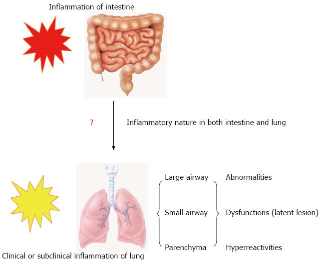
Figure 1 Model for inflammatory process in lung and intestine during inflammatory bowel disease.
Clinical or subclinical inflammation in small and large airways and the lung parenchyma accompanies the main inflammation in the bowel during inflammatory bowel disease.
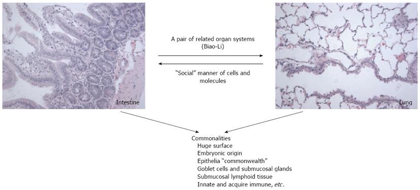
Figure 2 Summary model of plausible mechanisms of lung-intestine communication underlying inflammatory bowel disease-associated pulmonary involvement.
The lungs and intestines are a pair of mutually affected organs. The airways may intrinsically accompany inflammation of the bowel. This distal intrinsic inflammatory response may relate to the common features between the lung and intestine. According to Traditional Chinese Medicine, the lung and intestine have internal and external relationships (Biao-Li). This distal intrinsic inflammatory response may relate to the “social manner” of cells and molecules.
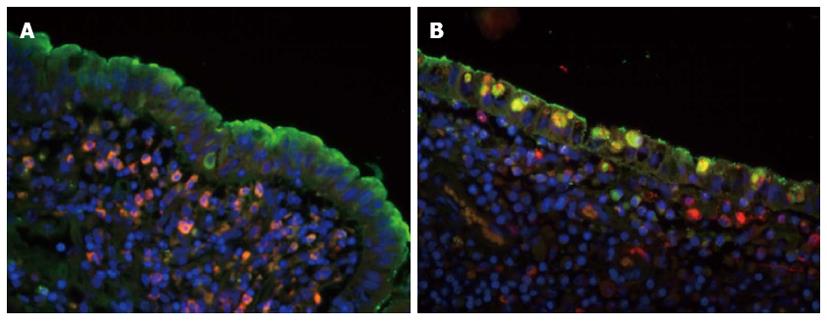
Figure 3 Co-localization of surfactant protein-A and CD68 in normal and inflammatory areas.
A: Surfactant protein (SP)-A -positive signal was indicated by fluorescence microscopy (green) fluorescence; CD68-positive signal was identified by rhodamine red-X (red) fluorescence; and the cell nucleus was indicated by Hoechst dye (blue). In the normal area, SP-A was located in the surface of the villi, specific epithelium and submucosae, lamina muscularis, mucosae and lymphoid tissues. CD68-positive cells were mainly found in the submucosae, lamina muscularis, mucosae, and lymphoid tissues and in the epithelium; B: In the inflammatory area, CD68-positive cells were dramatically increased in all levels of the bowel wall; especially CD68-positive macrophages in the epithelia of lamina mucosa. The SP-A-positive macrophages were recruited by activated epithelial cells. Double labeled SP-A and CD68 shows that some CD68-positive macrophages expressed SP-A like molecule in the inflammatory bowel disease tissues (original magnification, × 200). Figures reproduced with permission from reference [53].
- Citation: Wang H, Liu JS, Peng SH, Deng XY, Zhu DM, Javidiparsijani S, Wang GR, Li DQ, Li LX, Wang YC, Luo JM. Gut-lung crosstalk in pulmonary involvement with inflammatory bowel diseases. World J Gastroenterol 2013; 19(40): 6794-6804
- URL: https://www.wjgnet.com/1007-9327/full/v19/i40/6794.htm
- DOI: https://dx.doi.org/10.3748/wjg.v19.i40.6794


