Copyright
©2013 Baishideng Publishing Group Co.
World J Gastroenterol. Oct 7, 2013; 19(37): 6131-6143
Published online Oct 7, 2013. doi: 10.3748/wjg.v19.i37.6131
Published online Oct 7, 2013. doi: 10.3748/wjg.v19.i37.6131
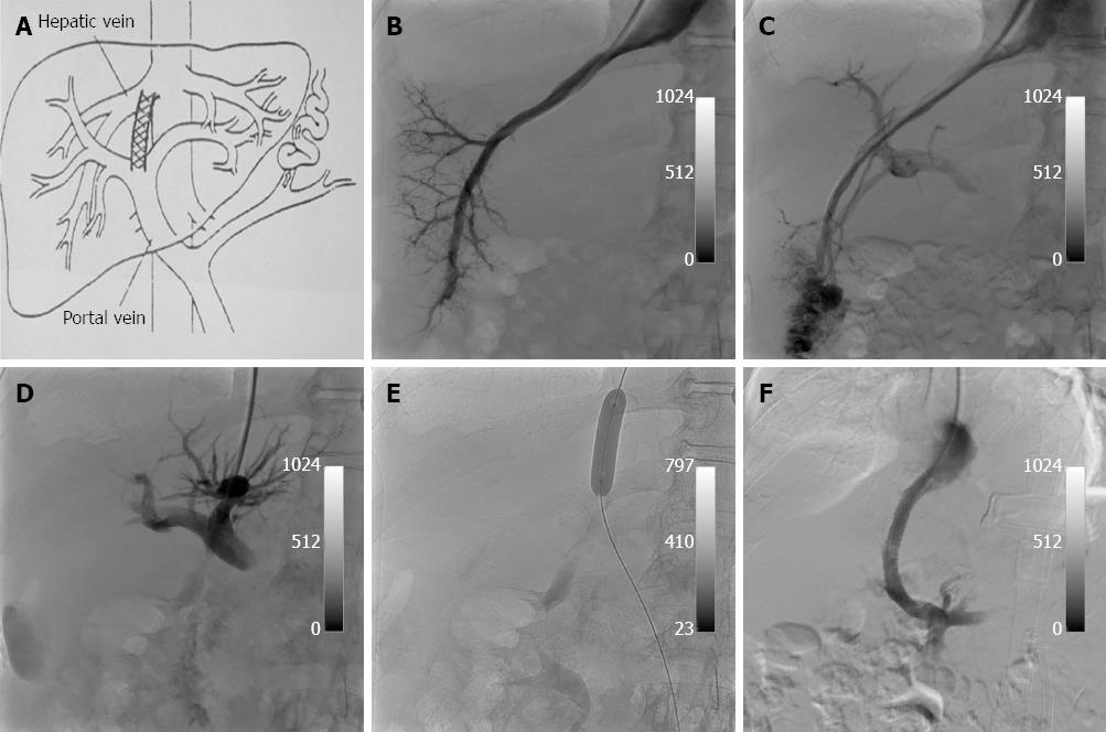
Figure 1 Conventional transjugular intrahepatic portosystemic shunt creation technique.
A: Schematic diagram shows transjugular intrahepatic portosystemic shunt (TIPS) connecting the right hepatic vein to the right portal vein. The shunt extends from main portal vein to confluence of right hepatic vein and inferior vena cava; B: Right hepatic venogram shows course of hepatic vein; C: Transhepatic portogram using iodinated contrast material shows course of portal veins; D: Injection of contrast medium through Colapinto needle confirms needle position within portal vein before passage of guidewire; E: Dilatation of a tract through the hepatic parenchyma that is interposed between the hepatic and portal veins; F: Portal venogram obtained after TIPS insertion shows flow through the FLUENCY polytetrafluoroethylene-covered stent. Peripheral portal vein branches are no longer opacified because of reversal of flow.
- Citation: Loffroy R, Estivalet L, Cherblanc V, Favelier S, Pottecher P, Hamza S, Minello A, Hillon P, Thouant P, Lefevre PH, Krausé D, Cercueil JP. Transjugular intrahepatic portosystemic shunt for the management of acute variceal hemorrhage. World J Gastroenterol 2013; 19(37): 6131-6143
- URL: https://www.wjgnet.com/1007-9327/full/v19/i37/6131.htm
- DOI: https://dx.doi.org/10.3748/wjg.v19.i37.6131


