Copyright
©2013 Baishideng Publishing Group Co.
World J Gastroenterol. Sep 21, 2013; 19(35): 5929-5932
Published online Sep 21, 2013. doi: 10.3748/wjg.v19.i35.5929
Published online Sep 21, 2013. doi: 10.3748/wjg.v19.i35.5929
Figure 1 Magnetic resonance imaging showing the liver lesion in segments III/IV.
Note the mass effect on the middle and left hepatic veins.
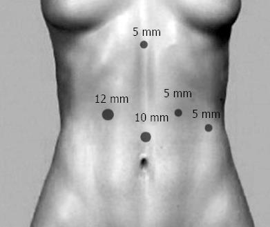
Figure 2 Patient positioning and trocar placement.
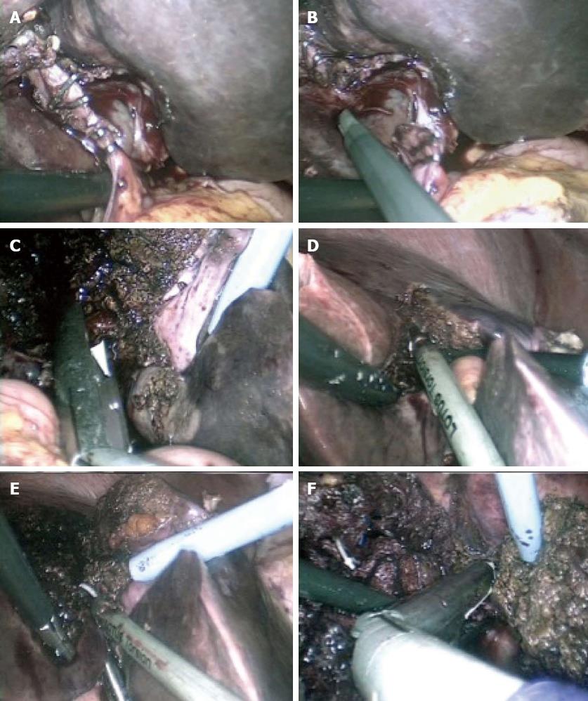
Figure 3 The operation.
A and B: Identification, dissection, and clip ligation of the replaced left hepatic artery; C: Dissection of the left portal vein; D and E: Parenchymal transection using the Lotus Ultrasonic Scalpel; F: Dissection of the left hepatic vein.
Figure 4 Left hepatectomy specimen (segments II-IV).
- Citation: Sotiropoulos GC, Stamopoulos P, Charalampoudis P, Molmenti EP, Voutsarakis A, Kouraklis G. Totally laparoscopic left hepatectomy using the Torsional Ultrasonic Scalpel. World J Gastroenterol 2013; 19(35): 5929-5932
- URL: https://www.wjgnet.com/1007-9327/full/v19/i35/5929.htm
- DOI: https://dx.doi.org/10.3748/wjg.v19.i35.5929


