Copyright
©2013 Baishideng Publishing Group Co.
World J Gastroenterol. Sep 7, 2013; 19(33): 5590-5592
Published online Sep 7, 2013. doi: 10.3748/wjg.v19.i33.5590
Published online Sep 7, 2013. doi: 10.3748/wjg.v19.i33.5590
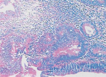
Figure 1 Pathological findings showed moderate-high grade dysplasia of the distal common bile duct lesion (hematoxylin/eosin staining, × 100).
Figure 2 Images taken via the trans-T-tube choledochoscope.
A: Before photodynamic treatment (PDT); B: Ten minutes after PDT; C: Two weeks after PDT; D: Three months after PDT.
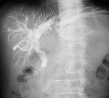
Figure 3 Trans-T-tube cholangiogram.
This image shows the dilated common bile duct and intrahepatic bile duct with an irregular filling defect in the distal common bile duct.
-
Citation: Zhou JJ, Xiong L, Li QL, Gu Y, Wen Y, Deng XF, Miao XY. Photodynamic therapy for high-grade dysplasia of bile duct
via a choledochoscope. World J Gastroenterol 2013; 19(33): 5590-5592 - URL: https://www.wjgnet.com/1007-9327/full/v19/i33/5590.htm
- DOI: https://dx.doi.org/10.3748/wjg.v19.i33.5590


