Copyright
©2013 Baishideng Publishing Group Co.
World J Gastroenterol. Aug 14, 2013; 19(30): 4907-4916
Published online Aug 14, 2013. doi: 10.3748/wjg.v19.i30.4907
Published online Aug 14, 2013. doi: 10.3748/wjg.v19.i30.4907
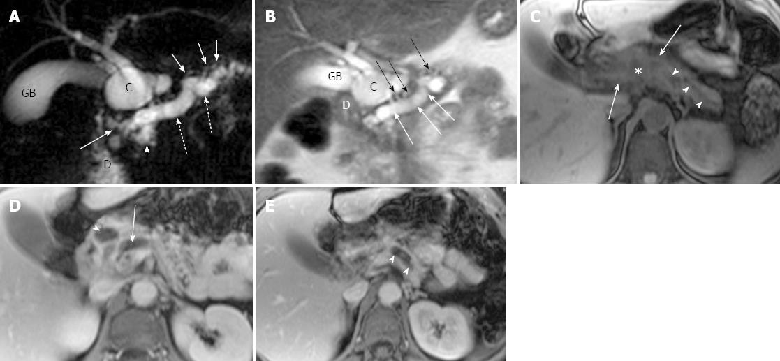
Figure 1 Magnetic resonance cholangiopancreatography and magnetic resonance imaging of recurrent acute pancreatitis involving the entire pancreas in a 44-year-old woman with several episodes of abdominal pain.
A: Coronal oblique thick-section rapid acquisition with relaxation enhancement magnetic resonance (RARE-MR) cholangiogram [infinite/1100 (effective), 40-mm section thickness] shows severe dilatation of pancreatic duct (dotted arrows) with stricture (solid arrow) just before entering the duodenum (D) and side branch ectasia (short arrows) as well. There is a pseudocyst (C) formation in pancreatic parenchyma. The gallbladder (GB) is distended and the common bile duct is dilated (arrowhead) as well; B: Thin-section half-Fourier RARE-MR cholangiogram [infinite/95 (effective), 3-mm section thickness] shows remarkable dilatation of dorsal pancreatic duct (white arrows) with severe side branch ectasia (black arrows). The pseudocyst (C) is formed in the pancreatic head region immediately adjacent to the duodenum (D) and GB; C: Axial precontrast T1WI SPGR shows the dilatation of pancreatic duct (arrowheads) and the pseudocyst (star) formation in the pancreatic neck were not easily appreciated. The signal intensity of the dorsal pancreas (arrows) is dramatically decreased; D: Axial postcontrast T1WI SPGR shows delayed enhancement of the dorsal pancreas and the wall of the pseudocysts (arrowhead). Dilatation of the pancreatic duct (arrow) was noted; E: Axial postcontrast T1WI SPGR shows delayed enhancement of the dorsal pancreas and the dilatation of the pancreatic duct (arrowheads).
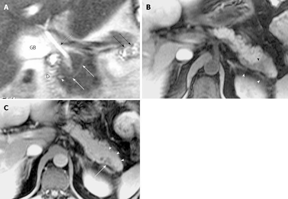
Figure 2 Magnetic resonance cholangiopancreatography and magnetic resonance imaging of recurrent acute pancreatitis with pancreas divisum only involving the pancreatic tail with a small santorinicele in a 44-year-old man with several episodes of abdominal pain.
A: Thin-section half-Fourier rapid acquisition with relaxation enhancement magnetic resonance (RARE- MR) cholangiogram [infinite/95 (effective), 3-mm section thickness] shows mild dilatation of the duct of Santorini (white arrows) with a focal enlargement consistent with santorinicele (arrowhead) at the entrance into the duodenum (D) via minor papilla. The side branch ectasia (black arrows) are noted in the pancreatic tail consistent with pancreatitis. Distended gallbladder (GB) and the CBD (dotted black arrow) are noted. B: Axial precontrast T1WI SPGR shows swelling and decrease of signal intensity in the pancreatic tail (black arrowhead), and thickening of the left anterior renal fascia (white arrowheads). C: Axial postcontrast T1WI SPGR shows mild delayed enhancement of pancreatic tail (arrowheads) with focal cystic changes (arrow).
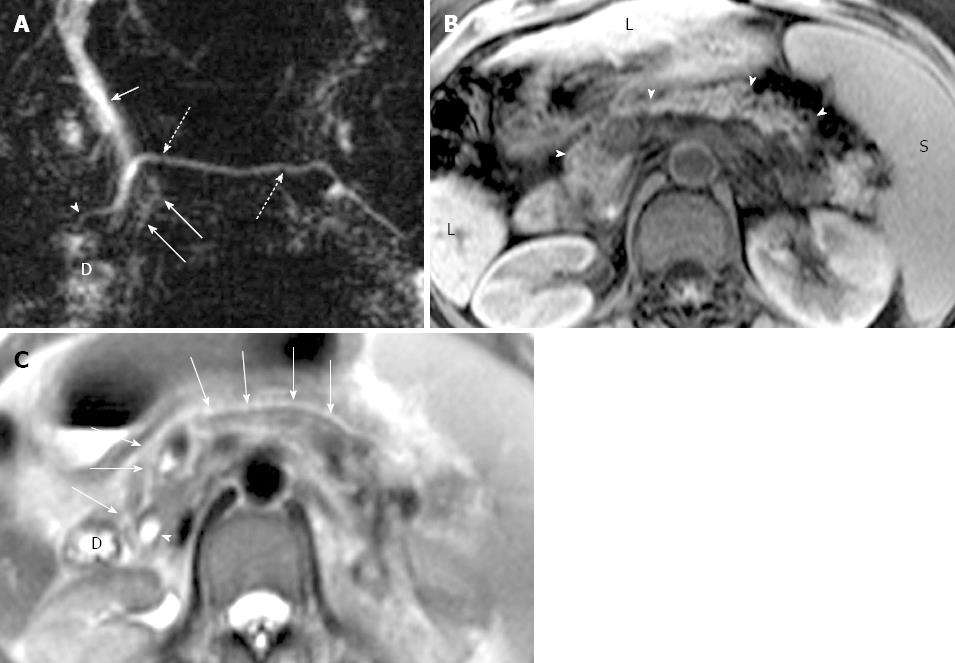
Figure 3 Magnetic resonance cholangiopancreatography and magnetic resonance imaging of incidental chronic pancreatitis in a 47-year-old woman with a history of liver cirrhosis.
A: Coronal oblique thick-section rapid acquisition with relaxation enhancement magnetic resonance (RARE-MR) cholangiogram [infinite/1100 (effective), 40-mm section thickness] of the pancreaticobiliary ducts shows slight dilatation with irregularity and strictures in the main pancreatic duct (dotted arrows) and in the duct of Santorini (arrowhead) just before entering the duodenum (D) via the minor papilla. Several side branch ectasia (long arrows) arising from the ventral duct and a normal common bile duct (short arrows) are demonstrated; B: Axial T1WI with fat saturation shows severe atrophy of the entire pancreas parenchyma (arrowheads). The liver (L) cirrhosis and splenomegaly (S) are noted; C: Axial T2WI shows main pancreatic duct (solid arrows) continued by duct of Santorini entering the duodenum (D) anterior to the common bile duct (arrowhead).
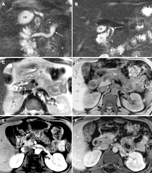
Figure 4 Magnetic resonance cholangiopancreatography and magnetic resonance imaging of recurrent acute pancreatitis only involving the dorsal pancreas in a 43-year-old man.
A: Coronal-oblique, thin-section half-Fourier rapid acquisition with relaxation enhancement magnetic resonance (RARE-MR) cholangiogram [infinite/95 (effective), 3-mm section thickness] shows marked dilatation (1 cm in diameter) of duct of Santorini (arrow) with apparent side branch ectasia (arrowheads). The gallbladder (GB) and the duodenum (D) are demonstrated clearly; B: Coronal-oblique, thin-section half-Fourier RARE MR cholangiogram [infinite/95 (effective), 3-mm section thickness] shows normal common bile duct (arrow) and ventral duct (arrowhead) of pancreas. The cystic changes secondary to pancreatitis in the pancreatic tail (dotted arrow) is shown while the GB and duodenum (D) appear normal; C: Axial T2WI shows dilatation and irregularity of duct of Santorini (short arrow) which enters the duodenum (D) via minor papilla. The ventral duct (solid arrow) and the pancreatic uncinate (arrowhead) are normal in size and signal intensity while the anterior portion of pancreatic head is abnormal with elevation of signal intensity; D: Axial precontrast T1WI SPGR shows that the pancreatic uncinate (arrowhead) is normal in size and signal intensity and the dorsal pancreas (DP) is abnormal with decreased T1 signal intensity and the swelling of the parenchyma; E: Axial T1WI SPGR after administration of Gd-DTPA at arterial phase shows normal enhancement of the pancreatic uncinate (arrowhead) and the enhancement of dorsal pancreas (PAN) is remarkably compromised with duct dilatation (arrow) and cystic changes; F: Axial T1WI SPGR at portal venous phase after administration of Gd-DTPA shows normal wash-out of contrast material in pancreatic uncinate (arrowhead) and delayed enhancement of the anterior portion of pancreatic head with dilatation of duct of Santorini (arrow).
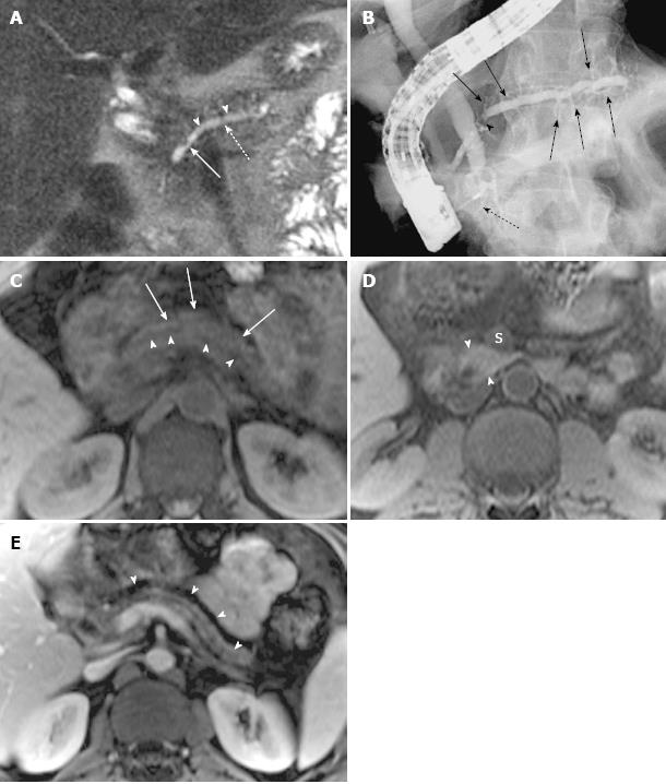
Figure 5 Magnetic resonance cholangiopancreatography and magnetic resonance imaging of recurrent acute pancreatitis only involving the dorsal pancreas, with endoscopic retrograde cholangiopancreatography correlation, in a 42-year-old man with several episodes of abdominal pain.
A: Coronal-oblique, thin-section half-Fourier rapid acquisition with relaxation enhancement magnetic resonance (RARE-MR) cholangiogram [infinite/95 (effective), 3-mm section thickness] shows remarkable dilatation of the dorsal pancreatic duct (dotted arrow) with a conspicuous stricture (solid arrow) and severe side branch ectasia (arrowheads); B: Endoscopic retrograde cholangiopancreatography shows the dilatation of dorsal pancreatic duct and duct of Santorini with a well-seen stricture (arrowhead) and remarkable side branch ectasia (arrows) continual and proximal to the minor papilla. The intrapancreatic segment of common bile duct (C) is very narrow while the other part of the CBD is dilated. The ventral duct is normal in size (dotted arrow); C: Axial precontrast T1WI SPGR shows a marked decrease of signal intensity of dorsal pancreas (arrows) with ductal dilatation (arrowheads); D: Axial precontrast T1WI SPGR shows that the uncinate of pancreas is normal in size and signal intensity (arrowheads) is much higher than the superior mesenteric vein (S) and similar to the liver; E: Axial postcontrast T1WI SPGR shows the atrophy of dorsal pancreas with delayed enhancement (arrowheads) and dilatation of the allied pancreatic duct.
- Citation: Wang DB, Yu J, Fulcher AS, Turner MA. Pancreatitis in patients with pancreas divisum: Imaging features at MRI and MRCP. World J Gastroenterol 2013; 19(30): 4907-4916
- URL: https://www.wjgnet.com/1007-9327/full/v19/i30/4907.htm
- DOI: https://dx.doi.org/10.3748/wjg.v19.i30.4907


