Copyright
©2013 Baishideng Publishing Group Co.
World J Gastroenterol. Jul 21, 2013; 19(27): 4422-4426
Published online Jul 21, 2013. doi: 10.3748/wjg.v19.i27.4422
Published online Jul 21, 2013. doi: 10.3748/wjg.v19.i27.4422
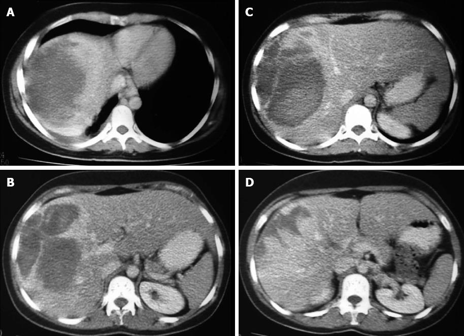
Figure 1 Unenhanced computed tomography scan showing liver injury with bleeding area, but also with areas of contrast enhancement.
A-D: The existence of bleeding denotes a hepatic laceration, but the presence of vascularization reinforces the presence of a focal lesion.
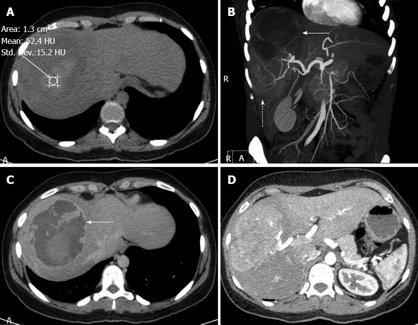
Figure 2 Computed tomography.
A: Unenhanced computed tomography (CT) scan showing the upper component of the lesion with a relatively high density of 52.4 UH, suggestive of bleeding; B: Contrast-enhanced CT in the coronal plane showing both components of the lesion. The upper portion (arrow) has a low density due to the absence of impregnation. The lower component (dotted arrow) is solid and hypervascular with internal calibrous arteries; C: Contrast-enhanced CT. The haematic component illustrates a contrast-enhanced capsule (arrow); D: Contrast-enhanced CT. The study after contrast in the arterial phase at the level of the solid component exhibits an area with greater permeability than the liver, suggestive of hypervascularity.
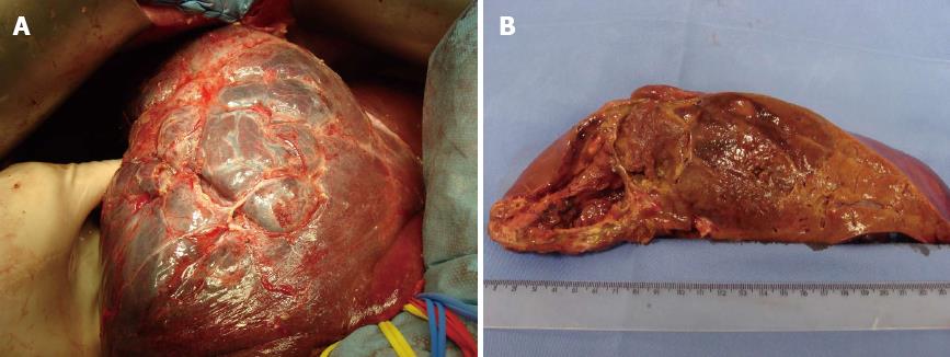
Figure 3 Entire right lobe of the liver.
A: Intraoperative image of the lesion. Mobilised liver after ligament release, demonstrating the heterogeneous mass occupying almost the entire right lobe of the liver (segments VI, VII and VIII), which was partially exophytic; B: Resected and sagittally cleaved surgical section at the transition between segments VII-VI and VIII-V. Note the adenomatous lesion with cavernomatous and haemorrhagic areas.
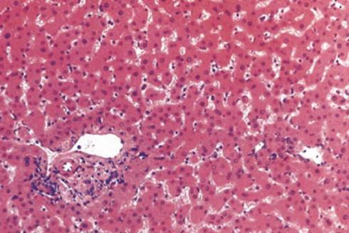
Figure 4 Histopathology showing the hepatocellular adenoma, a composed of cells that closely resemble normal hepatocytes, but in disorganized cords and with an abnormal lobular architecture (hematoxylin and eosin stain, × 200).
- Citation: Cotta-Pereira RL, Valente LF, De Paula DG, Eiras-Araújo AL, Iglesias AC. Rupture of a hepatic adenoma in a young woman after an abdominal trauma: A case report. World J Gastroenterol 2013; 19(27): 4422-4426
- URL: https://www.wjgnet.com/1007-9327/full/v19/i27/4422.htm
- DOI: https://dx.doi.org/10.3748/wjg.v19.i27.4422


