
















©2013 Baishideng Publishing Group Co.
World J Gastroenterol. Jul 21, 2013; 19(27): 4422-4426
Published online Jul 21, 2013. doi: 10.3748/wjg.v19.i27.4422
Published online Jul 21, 2013. doi: 10.3748/wjg.v19.i27.4422
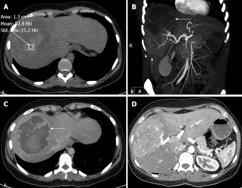
Figure 2 Computed tomography.
A: Unenhanced computed tomography (CT) scan showing the upper component of the lesion with a relatively high density of 52.4 UH, suggestive of bleeding; B: Contrast-enhanced CT in the coronal plane showing both components of the lesion. The upper portion (arrow) has a low density due to the absence of impregnation. The lower component (dotted arrow) is solid and hypervascular with internal calibrous arteries; C: Contrast-enhanced CT. The haematic component illustrates a contrast-enhanced capsule (arrow); D: Contrast-enhanced CT. The study after contrast in the arterial phase at the level of the solid component exhibits an area with greater permeability than the liver, suggestive of hypervascularity.
- Citation: Cotta-Pereira RL, Valente LF, De Paula DG, Eiras-Araújo AL, Iglesias AC. Rupture of a hepatic adenoma in a young woman after an abdominal trauma: A case report. World J Gastroenterol 2013; 19(27): 4422-4426
- URL: https://www.wjgnet.com/1007-9327/full/v19/i27/4422.htm
- DOI: https://dx.doi.org/10.3748/wjg.v19.i27.4422