Copyright
©2013 Baishideng Publishing Group Co.
World J Gastroenterol. Jul 21, 2013; 19(27): 4351-4355
Published online Jul 21, 2013. doi: 10.3748/wjg.v19.i27.4351
Published online Jul 21, 2013. doi: 10.3748/wjg.v19.i27.4351
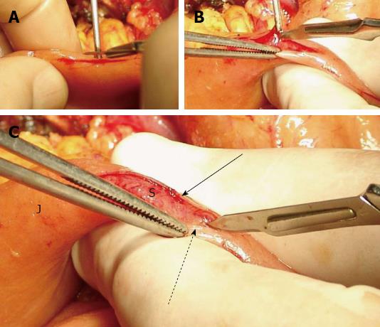
Figure 1 Preparation of the jejunal stump.
A: Incision on the jejunum; B: Seromuscular flap formation; C: The seromuscular layers are dissected free from the submucosa. J: Jejunum; S: Submucosa; Arrow: Posterior seromuscular flap; Dotted arrow: Anterior seromuscular flap.
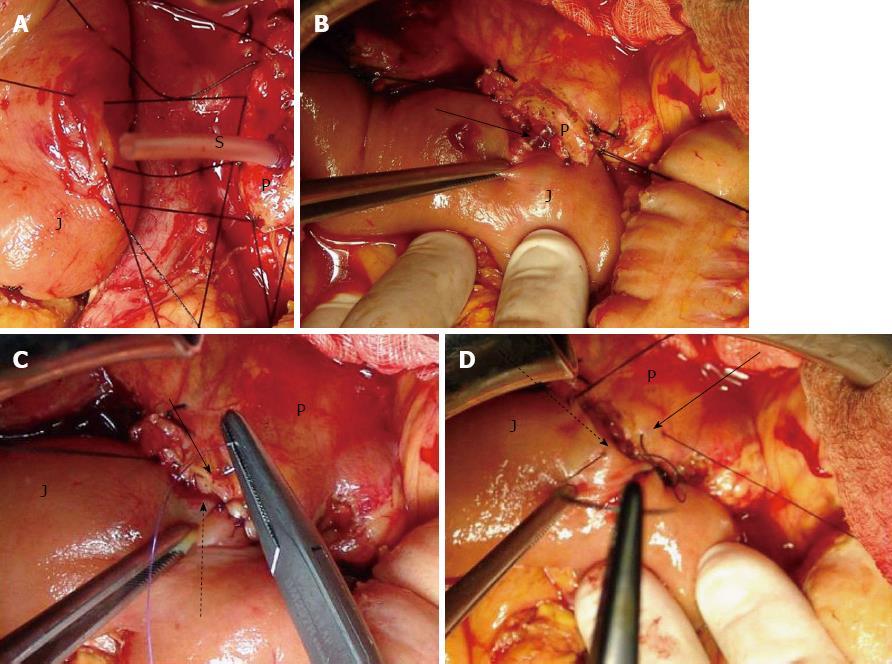
Figure 2 The posterior, anterior external layer of sutures and stent in place.
A: The posterior (dorsal) external layer of sutures; B: Stent in place. Arrow: The stent enters the jejunal small opening, which has been created in the middle of the dissected submucosal surface; C: The anterior (ventral) internal layer of sutures. Arrow: Anterior pancreatic cut edge border; Dotted arrow: Anterior seromuscular flap; D: The anterior (ventral) external layer of sutures. Arrow: Anterior pancreatic part of the capsular parenchyma; Dotted arrow: Anterior part of the jejunal seromuscular layer. J: Jejunum; S: Stent; P: Pancreatic stump cut surface.
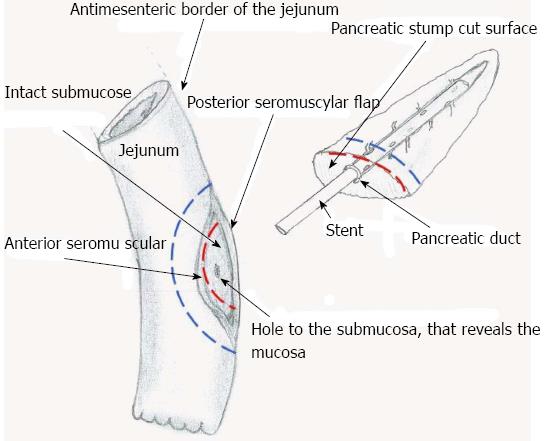
Figure 3 This is a schema that shows the dissected surface of the jejunum and the cut surface of the pancreas that are approximated.
The red dotted line represents the ventral internal suturing layer (i.e., the 3rd one). The blue dotted line represents the ventral external suturing layer (i.e., the 4th one).
- Citation: Katsaragakis S, Larentzakis A, Panousopoulos SG, Toutouzas KG, Theodorou D, Stergiopoulos S, Androulakis G. A new pancreaticojejunostomy technique: A battle against postoperative pancreatic fistula. World J Gastroenterol 2013; 19(27): 4351-4355
- URL: https://www.wjgnet.com/1007-9327/full/v19/i27/4351.htm
- DOI: https://dx.doi.org/10.3748/wjg.v19.i27.4351


