Copyright
©2013 Baishideng Publishing Group Co.
World J Gastroenterol. Jul 21, 2013; 19(27): 4351-4355
Published online Jul 21, 2013. doi: 10.3748/wjg.v19.i27.4351
Published online Jul 21, 2013. doi: 10.3748/wjg.v19.i27.4351
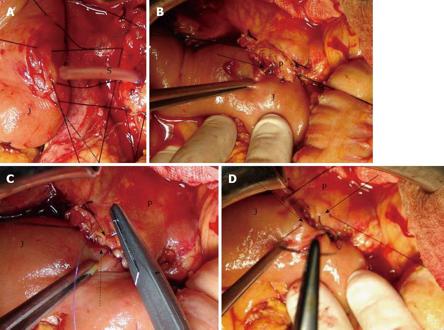
Figure 2 The posterior, anterior external layer of sutures and stent in place.
A: The posterior (dorsal) external layer of sutures; B: Stent in place. Arrow: The stent enters the jejunal small opening, which has been created in the middle of the dissected submucosal surface; C: The anterior (ventral) internal layer of sutures. Arrow: Anterior pancreatic cut edge border; Dotted arrow: Anterior seromuscular flap; D: The anterior (ventral) external layer of sutures. Arrow: Anterior pancreatic part of the capsular parenchyma; Dotted arrow: Anterior part of the jejunal seromuscular layer. J: Jejunum; S: Stent; P: Pancreatic stump cut surface.
- Citation: Katsaragakis S, Larentzakis A, Panousopoulos SG, Toutouzas KG, Theodorou D, Stergiopoulos S, Androulakis G. A new pancreaticojejunostomy technique: A battle against postoperative pancreatic fistula. World J Gastroenterol 2013; 19(27): 4351-4355
- URL: https://www.wjgnet.com/1007-9327/full/v19/i27/4351.htm
- DOI: https://dx.doi.org/10.3748/wjg.v19.i27.4351


