Copyright
©2013 Baishideng Publishing Group Co.
World J Gastroenterol. Jun 28, 2013; 19(24): 3792-3801
Published online Jun 28, 2013. doi: 10.3748/wjg.v19.i24.3792
Published online Jun 28, 2013. doi: 10.3748/wjg.v19.i24.3792
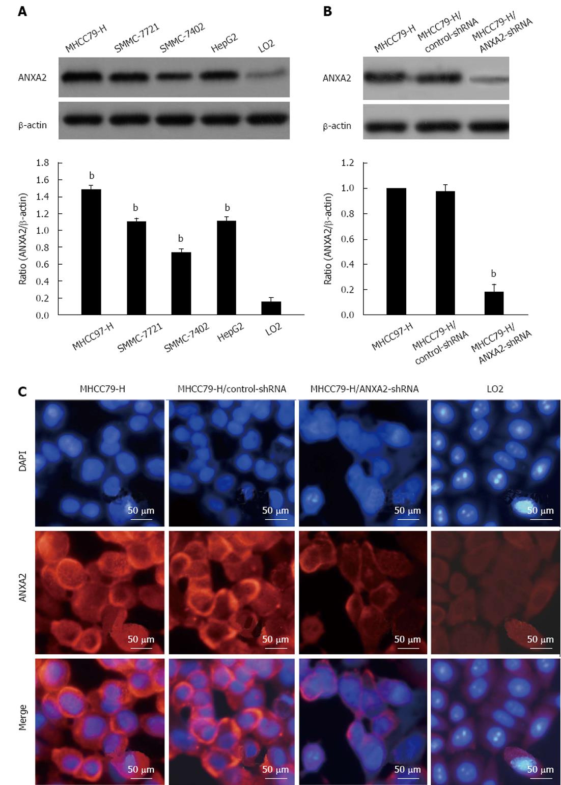
Figure 1 Annexin A2 expression level in hepatoma cells and silencing efficiency of small hairpin RNA in MHCC97-H cells.
A: Representative Western blotting images of hepatocellular carcinoma cell lines and the normal hepatic cell line LO2. bP < 0.01 vs LO2; B: Representative Western blotting images of annexin A2 (ANXA2) silencing upon transfection of small hairpin RNA (shRNA). bP < 0.01 vs MHCC97-H; C: Representative immunofluorescence images of ANXA2 cellular distribution (× 400). DAPI: 4,6-diamidino-2-phenylindole.
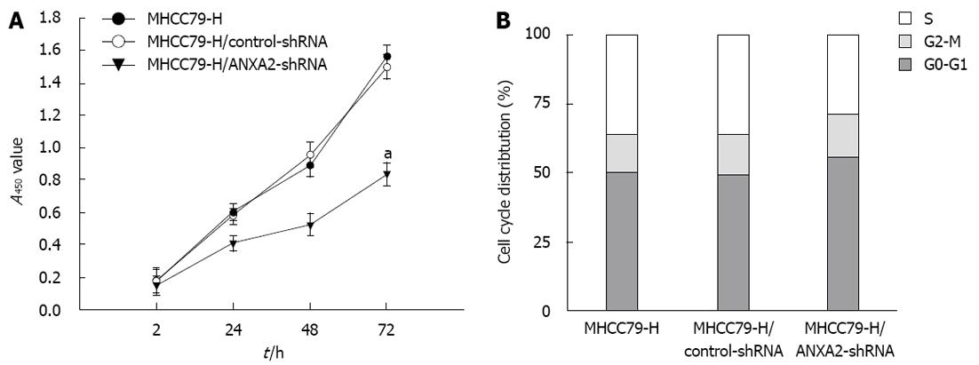
Figure 2 Effect of small hairpin RNA-mediated annexin A2 silencing on proliferation and cell cycling of MHCC97-H cells.
A: Cellular proliferation assay. aP < 0.05 vs the A450 of MHCC97-H cells at 72 h; B: Cell cycle assay. P < 0.05 for percentage of MHCC97-H/annexin A2 (ANXA2)-small hairpin RNA (shRNA) cells in S phase vs that of MHCC97-H cells.
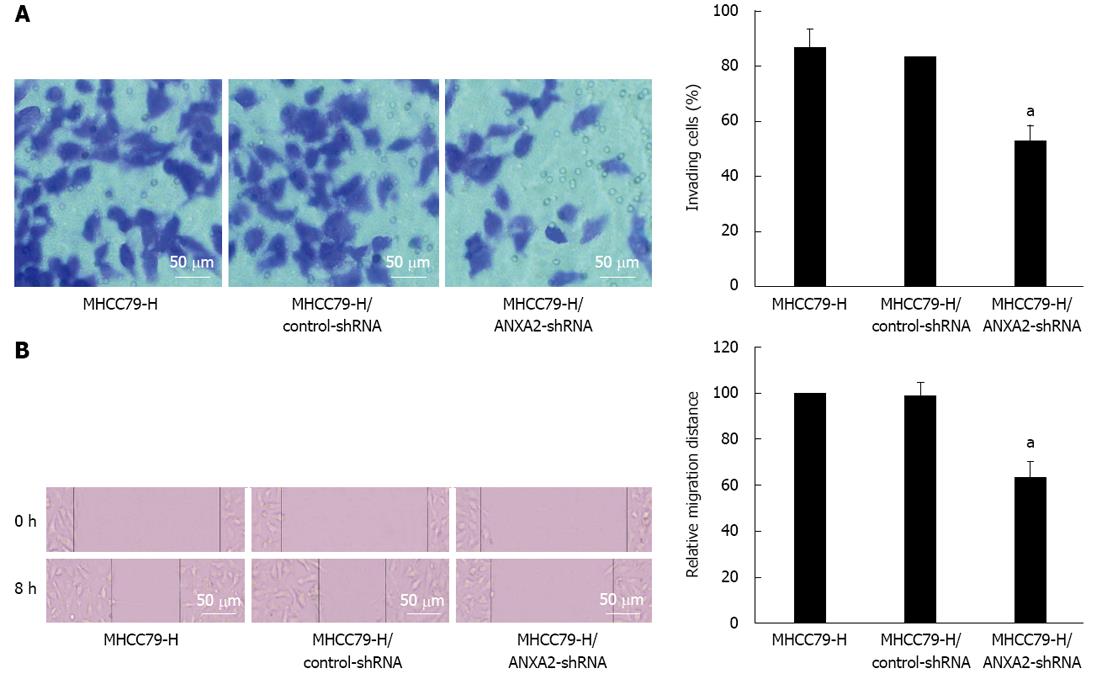
Figure 3 Suppressive effect of small hairpin RNA -mediated annexin A2 silencing on the invasion and migration potential of MHCC97-H cells.
A: Representative images of invasive cells (stained with crystal violet) from: the MHCC97-H group; the MHCC97-H/control-small hairpin RNA (shRNA) group; the MHCC97-H/annexin A2 (ANXA2)-small hairpin RNA (shRNA) group; B: Representative images of cell migration. aP < 0.05 vs MHCC97-H cells.
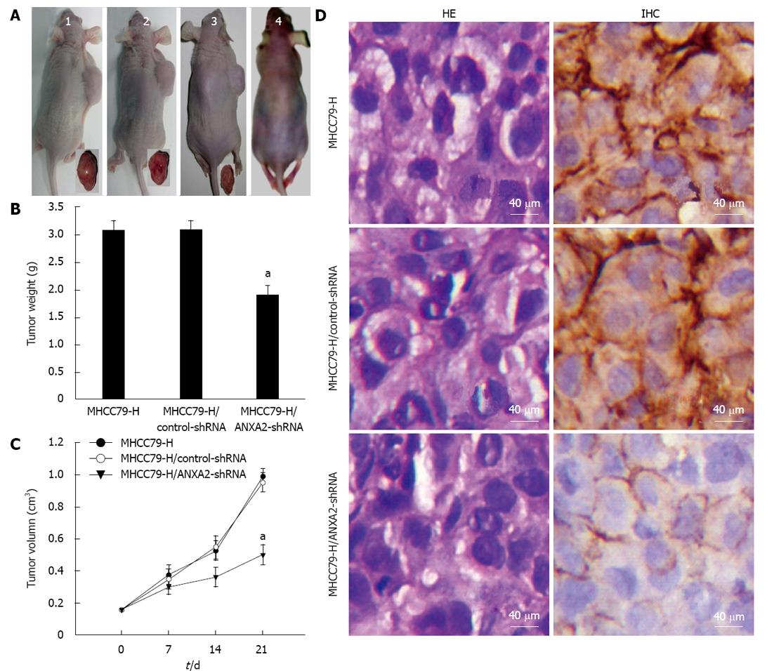
Figure 4 Inhibitive effect of small hairpin RNA-mediated annexin A2 silencing on xenograft tumour growth in vivo.
A: Representative images of xenografted and control mice and resected tumours. The MHCC97-H (untransfected) group (1); the MHCC97-H/control-small hairpin RNA (shRNA) group (2); the MHCC97-H/annexin A2 (ANXA2)-shRNA group (3); the blank control group (4). Tumorigenic nude mice appeared obviously emaciated, especially the those in the MHCC97-H group and MHCC97-H/control-shRNA group; B: Average tumour weights. aP < 0.05 vs MHCC97-H group; C: Tumour growth rates. aP < 0.05 vs MHCC97-H group at post-injection day 21; D: Representative immunohistochemical analysis and hematoxylin and eosin staining results (× 400). The density of ANXA2 staining (brown) in the cytoplasm of MHCC97-H/ANXA2-shRNA cells was obviously lower than that for the MHCC97-H cells or the MHCC97-H/control-shRNA cells. ANXA2 was mainly localized in the membrane of the MHCC97-H/ANXA2-shRNA cells, and localized in both the membrane and cytoplasm of the MHCC97-H cells and the MHCC97-H/control-shRNA cells. The morphological characteristics of subcutaneous xenograft tumours derived from MHCC97-H/ANXA2-shRNA cells were not fundamentally different from the other tumours.
- Citation: Zhang HJ, Yao DF, Yao M, Huang H, Wang L, Yan MJ, Yan XD, Gu X, Wu W, Lu SL. Annexin A2 silencing inhibits invasion, migration, and tumorigenic potential of hepatoma cells. World J Gastroenterol 2013; 19(24): 3792-3801
- URL: https://www.wjgnet.com/1007-9327/full/v19/i24/3792.htm
- DOI: https://dx.doi.org/10.3748/wjg.v19.i24.3792


