Copyright
©2013 Baishideng Publishing Group Co.
World J Gastroenterol. Jun 14, 2013; 19(22): 3512-3516
Published online Jun 14, 2013. doi: 10.3748/wjg.v19.i22.3512
Published online Jun 14, 2013. doi: 10.3748/wjg.v19.i22.3512
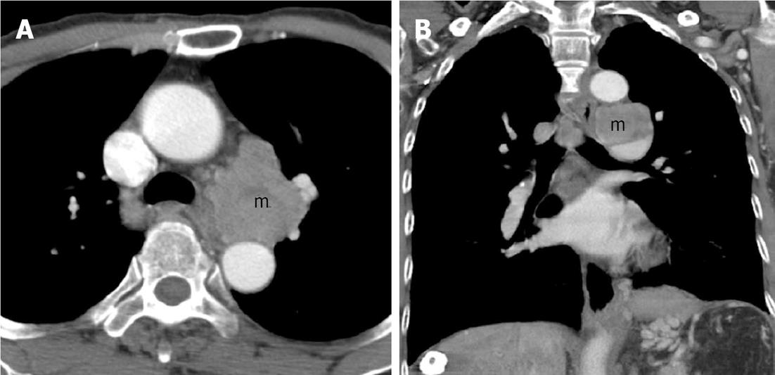
Figure 1 A chest computer tomography scan of the aortopulmonary window of the mediastinum revealed a 5 cm metastatic lymphadenopathy (m).
A: Transverse view; B: Sagittal view.
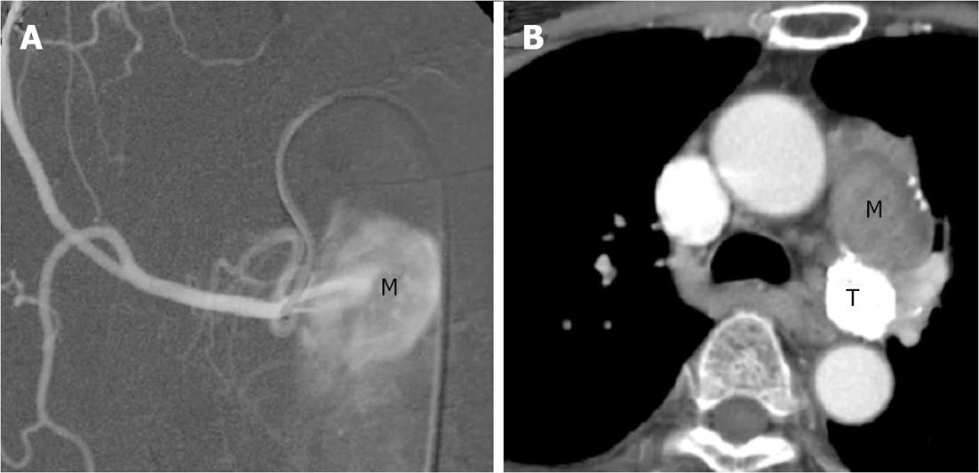
Figure 2 First session of transarterial embolization for mediastinal tumor.
A: An angiogram of the right superior bronchial arterial branch showed a metastatic hepatocellular carcinoma (M) in the left hilum supplying by the first branch of right superior bronchial artery. Embolization was performed by lipiodol only; B: Chest computer tomography (CT) scan: Lipiodol retention in posterior portion of the mass (T) over aortopulmonary window region and decreased in size, but the anterior portion of the mass (M) over aortopulmonary window region increased in size as compared with previous CT.
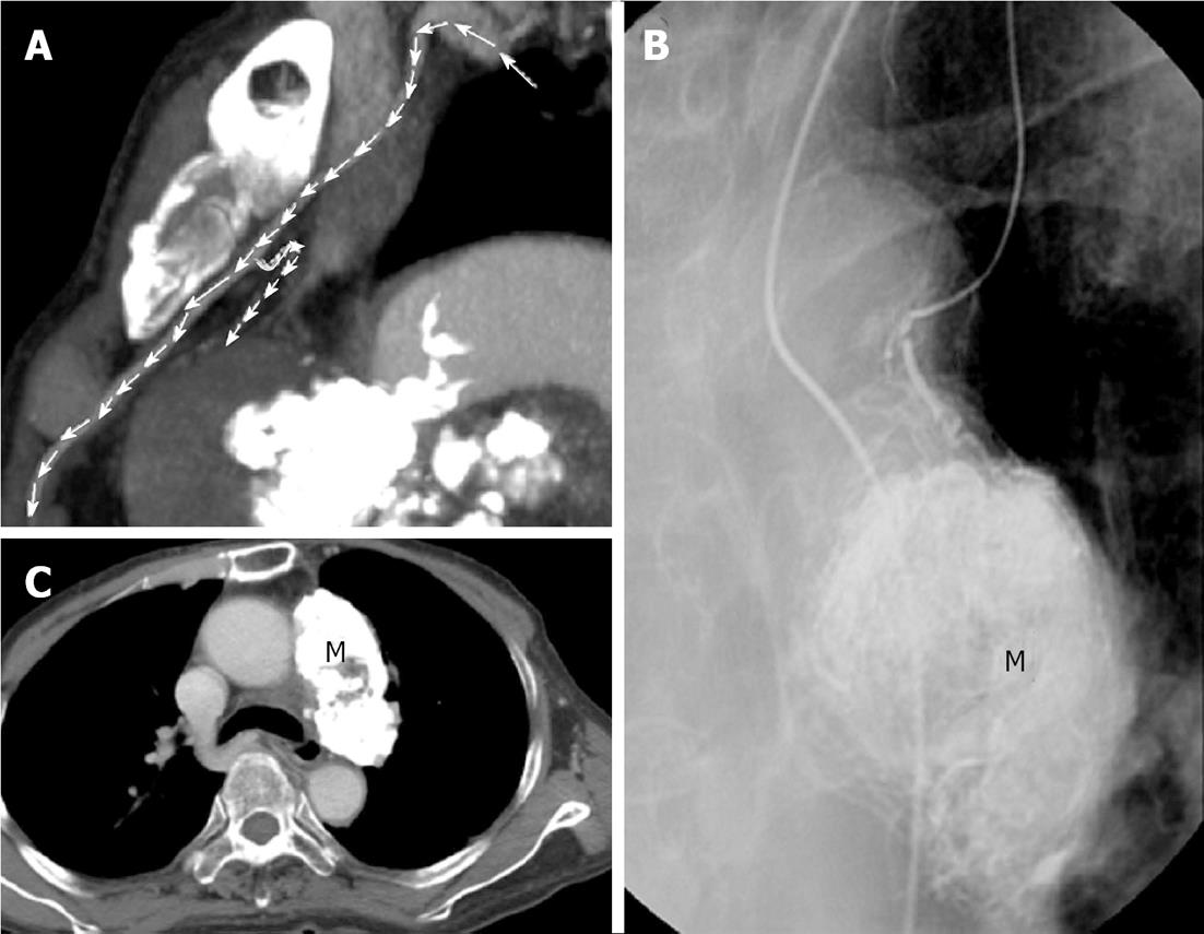
Figure 3 Successful control of mediastinal metastasis via selection of the supplying vessel.
A: Multi-detector row computed tomography showed a supplying artery from small branch of left internal mammary artery to residual tumor (arrows); B: Tumor supplying artery of left internal thoracic (mammary) artery was selected via a 3 Fr microcatheter. Transarterial embolization for tumor (M) was done by using 6 mL of lipiodol; C: Follow-up chest computed tomography showed the metastatic tumor (M) in left anterior mediastinum was well embolized.
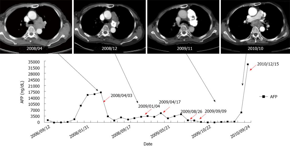
Figure 4 Alpha-fetoprotein levels in time course of treatment.
Every session of transarterial embolization is shown with a red arrow. The computed tomography (CT) images represented the progression of mediastinal tumor and treatment response of transarterial embolization. The CT image on the far right was taken about three months before death of the patient. The tumor compressed the left bronchus, which may explain the cause of the patient’s pneumonia.
- Citation: Chen CC, Yeh HZ, Chang CS, Ko CW, Lien HC, Wu CY, Hung SW. Transarterial embolization of metastatic mediastinal hepatocellular carcinoma. World J Gastroenterol 2013; 19(22): 3512-3516
- URL: https://www.wjgnet.com/1007-9327/full/v19/i22/3512.htm
- DOI: https://dx.doi.org/10.3748/wjg.v19.i22.3512


