Copyright
©2013 Baishideng Publishing Group Co.
World J Gastroenterol. May 28, 2013; 19(20): 3165-3168
Published online May 28, 2013. doi: 10.3748/wjg.v19.i20.3165
Published online May 28, 2013. doi: 10.3748/wjg.v19.i20.3165
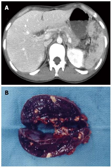
Figure 1 Contrast-enhanced abdominal computed tomography scan showing multiple spleen abscesses (A), and macroscopic findings of the cut surface of the resected spleen (B).
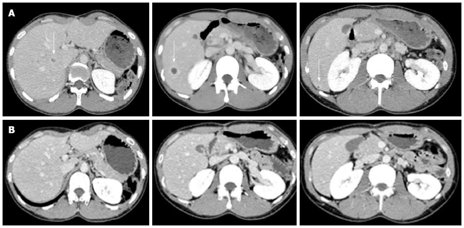
Figure 2 Contrast-enhanced abdominal computed tomography scan showing multiple liver abscesses (arrow) before (A) and 4 wk after corticosteroid therapy (B).
Multiple areas of low-attenuation with ring enhancement were seen in the liver as indicated by the arrows (A). However, the areas vanished after treatment (B).
- Citation: Maeshima K, Ishii K, Inoue M, Himeno K, Seike M. Behçet’s disease complicated by multiple aseptic abscesses of the liver and spleen. World J Gastroenterol 2013; 19(20): 3165-3168
- URL: https://www.wjgnet.com/1007-9327/full/v19/i20/3165.htm
- DOI: https://dx.doi.org/10.3748/wjg.v19.i20.3165


