Copyright
©2013 Baishideng Publishing Group Co.
World J Gastroenterol. Jan 14, 2013; 19(2): 311-315
Published online Jan 14, 2013. doi: 10.3748/wjg.v19.i2.311
Published online Jan 14, 2013. doi: 10.3748/wjg.v19.i2.311
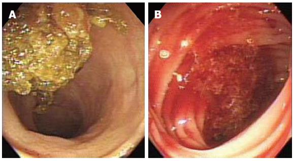
Figure 1 Initial colonoscopy.
A: The terminal ileum showing yellowish-colored stool with no evidence of upper gastrointestinal bleeding; B: The ascending colon showing fresh blood without an observable source.
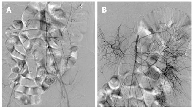
Figure 2 Mesenteric artery angiography.
A: The inferior mesenteric artery angiography showing no source of bleeding; B: The superior mesenteric artery angiography showing no source of bleeding.
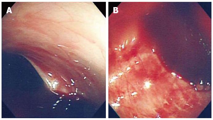
Figure 3 The second colonoscopy.
A: Massive hemorrhage within the proximal ascending colon without a definite bleeding focus; B: Fresh blood within the distal ascending colon without a definite bleeding focus.
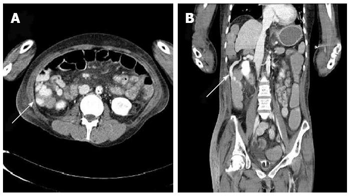
Figure 4 Abdominal computed tomography.
A: Portal phase axial view with white arrow showing the varix surrounding and protruding inside the ascending colon formed by portacaval shunt; B: Coronal view with white arrow showing the colon varix.
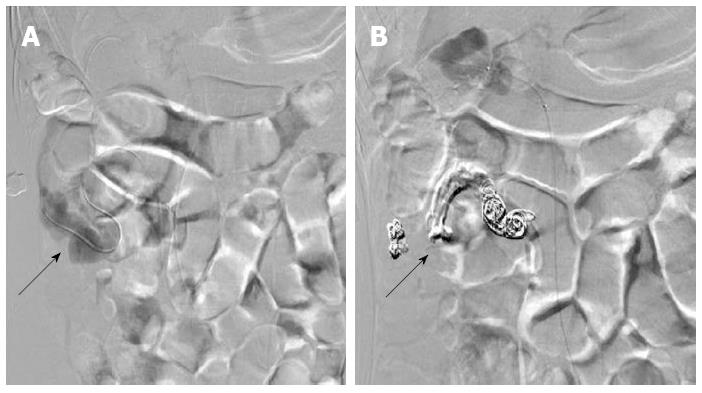
Figure 5 Venous coil embolization.
A: Venography showing the varix within the ascending colon (black arrow); B: Coil embolization (black arrow) and histoacryl injection was performed to obliterate the varix.
- Citation: Ko BS, Kim WT, Chang SS, Kim EH, Lee SW, Park WS, Kim YS, Nam SW, Lee DS, Kim JC, Kang SB. A case of ascending colon variceal bleeding treated with venous coil embolization. World J Gastroenterol 2013; 19(2): 311-315
- URL: https://www.wjgnet.com/1007-9327/full/v19/i2/311.htm
- DOI: https://dx.doi.org/10.3748/wjg.v19.i2.311


