Copyright
©2013 Baishideng Publishing Group Co.
World J Gastroenterol. May 21, 2013; 19(19): 2974-2978
Published online May 21, 2013. doi: 10.3748/wjg.v19.i19.2974
Published online May 21, 2013. doi: 10.3748/wjg.v19.i19.2974
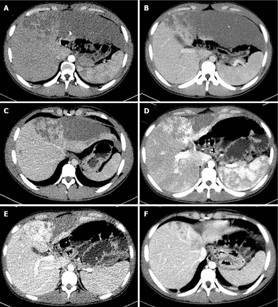
Figure 1 Axial images of multi-detector computed tomography.
Multi-detector computed tomography (MDCT) images at the first visit (A-C), and corresponding MDCT slices just before the operation (i.e., one month after transcatheter arterial embolization) (E-G).
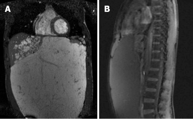
Figure 2 Coronal (A) and sagittal (B) views of the hemangioma from magnetic resonance imaging.
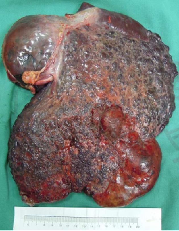
Figure 3 Intra-operative photograph of the tumor.
- Citation: Zhou JX, Huang JW, Wu H, Zeng Y. Successful liver resection in a giant hemangioma with intestinal obstruction after embolization. World J Gastroenterol 2013; 19(19): 2974-2978
- URL: https://www.wjgnet.com/1007-9327/full/v19/i19/2974.htm
- DOI: https://dx.doi.org/10.3748/wjg.v19.i19.2974


