Copyright
©2013 Baishideng Publishing Group Co.
World J Gastroenterol. May 14, 2013; 19(18): 2830-2834
Published online May 14, 2013. doi: 10.3748/wjg.v19.i18.2830
Published online May 14, 2013. doi: 10.3748/wjg.v19.i18.2830
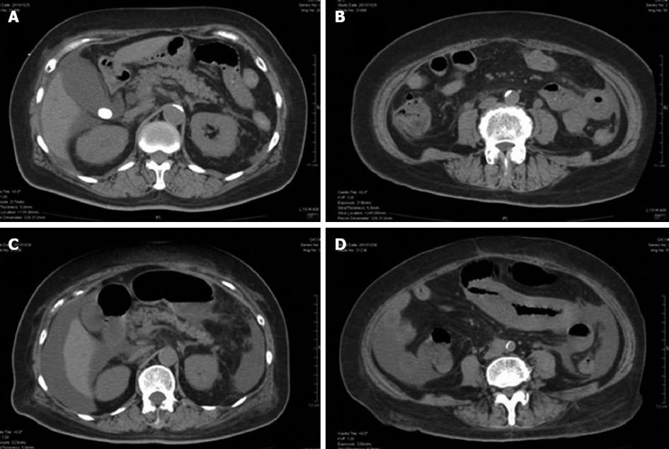
Figure 1 Abdominal computed tomography findings.
A, B: On day 1 showed ascites and bowel wall thickening; C, D: On day 6, both of the ascites and the bowel wall thickening, especially jejunum, had severely worsened.
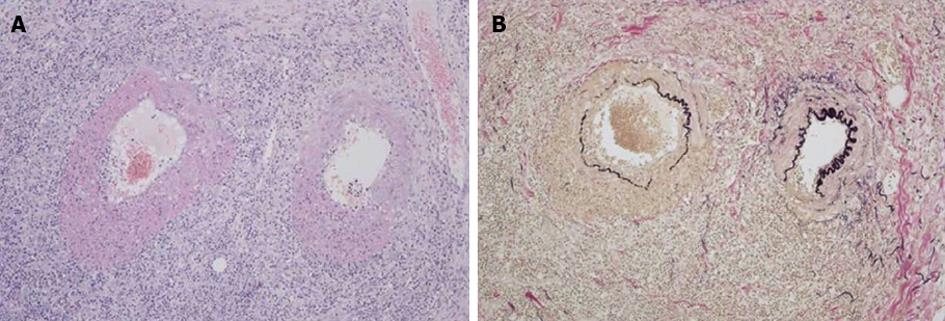
Figure 2 Pathological specimen from the resected jejunum showing fibrinoid necrosis of the arterial wall and infiltration of inflammatory cells around the arteries.
A: Hematoxylin and eosin staining. B: Elastica van Gieson staining. Original magnification ×100.
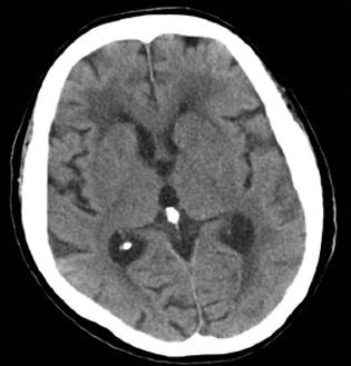
Figure 3 Head computed tomography, which revealed no clear evidence consistent with left hemiparesis.
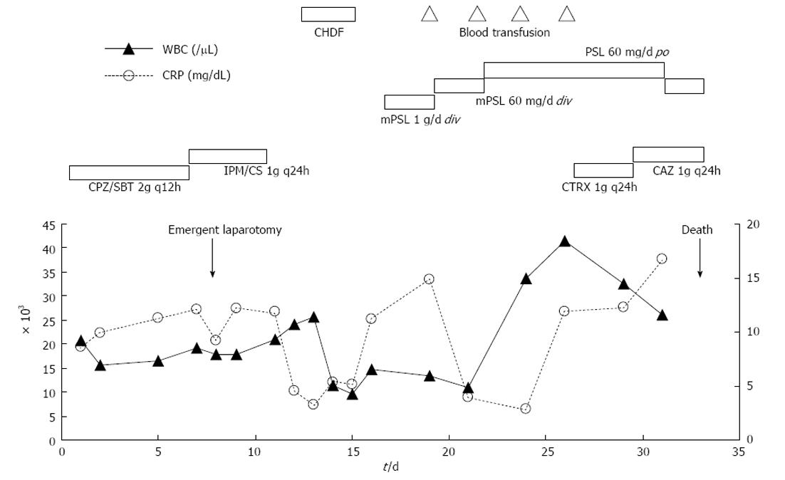
Figure 4 Clinical course of the patient.
Cefoperazone/sulbactam (CPZ/SBT) was started at day 1 and switched to imipenem/cilastatin (IPM/CS) at day 7. Ceftriaxone (CTRX) was started at day 26 and switched to ceftazidime (CAZ) at day 29. Steroid pulse therapy [methylprednisolone (mPSL) 1 g/d for 3 d] was started at day 16, and changed to maintenance dose of mPSL (60 mg/d) at day 19, then that was switched to oral steroid (PSL 60 mg/d) at day 22. According to deterioration of general status, steroid was again administered intravenously since day 31. Continuous hemodiafiltration (CHDF) was performed from day 13 to day 15, and intermittent blood transfusion was done at day 19, 21, 23 and 26. WBC: White blood cell; CRP: C-reactive protein.
- Citation: Hiraike Y, Kodaira M, Sano M, Terazawa Y, Yamagata S, Terada S, Ohura M, Kuriki K. Polyarteritis nodosa diagnosed by surgically resected jejunal necrosis following acute abdomen. World J Gastroenterol 2013; 19(18): 2830-2834
- URL: https://www.wjgnet.com/1007-9327/full/v19/i18/2830.htm
- DOI: https://dx.doi.org/10.3748/wjg.v19.i18.2830


