Copyright
©2013 Baishideng Publishing Group Co.
World J Gastroenterol. Apr 28, 2013; 19(16): 2555-2559
Published online Apr 28, 2013. doi: 10.3748/wjg.v19.i16.2555
Published online Apr 28, 2013. doi: 10.3748/wjg.v19.i16.2555
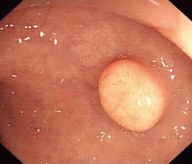
Figure 1 Endoscopic findings of rectal carcinoids.
Endoscopy shows yellow or gray submucosal nodules, which are hard and covered by normal-appearing mucosa.
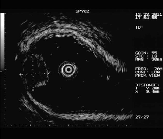
Figure 2 Rectal carcinoids under micro-probe ultrasound.
The hypoechoic lesions are mainly derived from submucosa or muscularis mucosa.
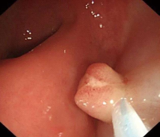
Figure 3 Endoscopic loop ligation after injection.
Figure 4 Endoscopic resection of the lesions.
The whole rectal carcinoid mass is visible in the endoscopically resected lesion.
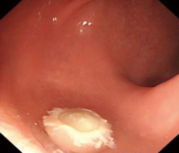
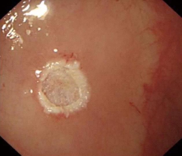
Figure 5 Wounds after endoscopic resection.
After the endoscopic resection of the rectal carcinoid, the wound is complete and no residual mass is present.
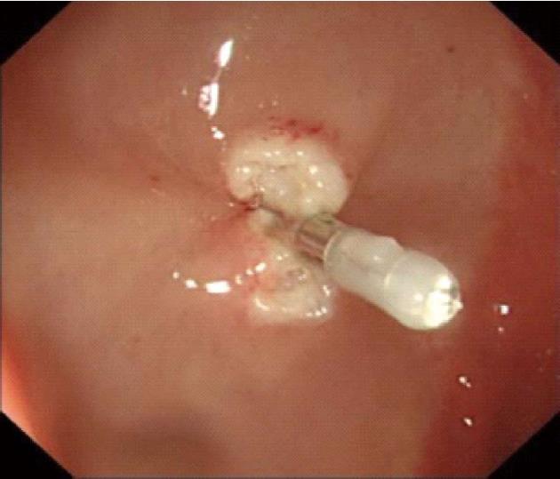
Figure 6 Wounds after endoscopic clipping.
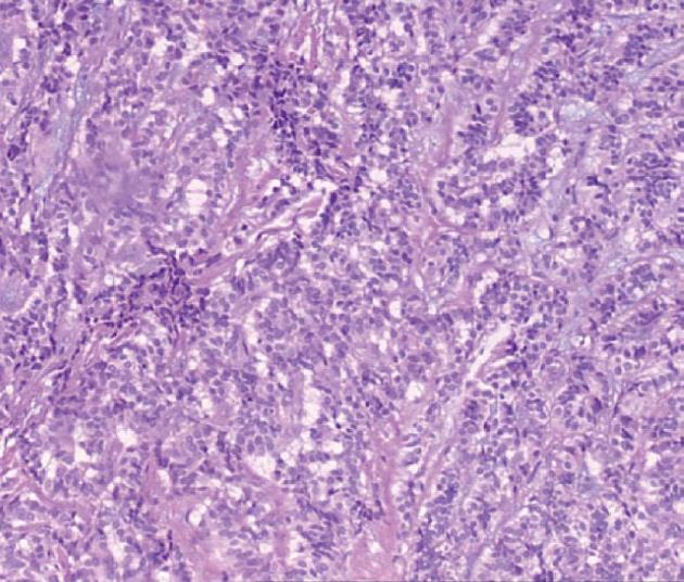
Figure 7 Pathological manifestations of rectal carcinoids.
The carcinoid cells are small oval or polygonal, nested or glandular-like, and with a regular and small nucleus under microscope (magnification × 400).
- Citation: Zhou FR, Huang LY, Wu CR. Endoscopic mucosal resection for rectal carcinoids under micro-probe ultrasound guidance. World J Gastroenterol 2013; 19(16): 2555-2559
- URL: https://www.wjgnet.com/1007-9327/full/v19/i16/2555.htm
- DOI: https://dx.doi.org/10.3748/wjg.v19.i16.2555


