Copyright
©2013 Baishideng Publishing Group Co.
World J Gastroenterol. Apr 21, 2013; 19(15): 2437-2440
Published online Apr 21, 2013. doi: 10.3748/wjg.v19.i15.2437
Published online Apr 21, 2013. doi: 10.3748/wjg.v19.i15.2437
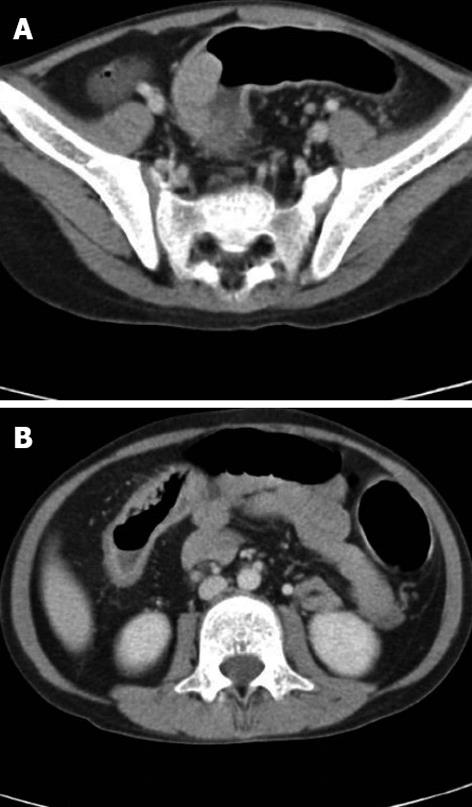
Figure 1 Preoperative computed tomography.
A: Enhanced protruding polypoid mass at the sigmoid colon; B: diffuse colonic wall thickening with loss of haustra (“lead pipe appearance”).
Figure 2 Colonoscopy findings.
A: Diffuse granular lesions; B: Multiple ulcerations from the transverse colon to the rectum; C: A polypoid mass at the sigmoid colon.
Figure 3 Gross appearance of the colon.
A: There was a 5 cm × 3 cm-sized polypoid mass at the sigmoid colon; B: There was severe nodularity with fibrosis in the whole colon.
- Citation: Noh SY, Oh SY, Kim SH, Kim HY, Jung SE, Park KW. Fifteen-year-old colon cancer patient with a 10-year history of ulcerative colitis. World J Gastroenterol 2013; 19(15): 2437-2440
- URL: https://www.wjgnet.com/1007-9327/full/v19/i15/2437.htm
- DOI: https://dx.doi.org/10.3748/wjg.v19.i15.2437


