Copyright
©2013 Baishideng Publishing Group Co.
World J Gastroenterol. Apr 21, 2013; 19(15): 2433-2436
Published online Apr 21, 2013. doi: 10.3748/wjg.v19.i15.2433
Published online Apr 21, 2013. doi: 10.3748/wjg.v19.i15.2433
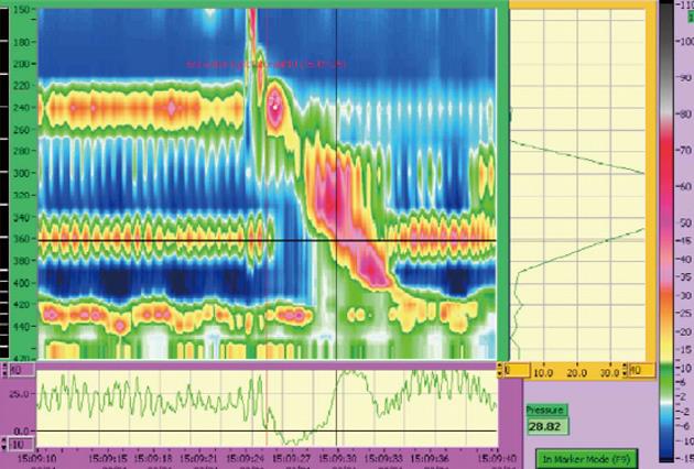
Figure 1 Manometry study demonstrating a high pressure band in the lower oesophagus (at the 360 mm mark/36 cm) consistent with arterial pulsations.
This pressure band does not manometrically cause obstruction with a clear relaxation across the region of interest during the swallow.
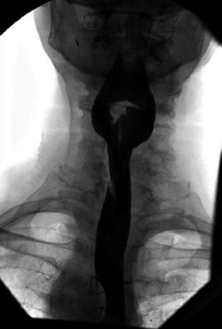
Figure 2 Barium oesophogram demonstrating the extrinsic impression on the oesophagus secondary to the aberrant left subclavian artery superiorly and right aortic arch distally.
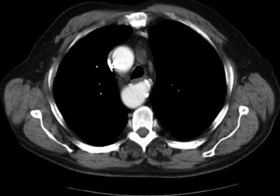
Figure 3 Computed tomography chest demonstrating the right aortic arch and dilatation at the origin of the aberrant left subclavian artery (Komerrell’s diverticulum).
- Citation: Bennett AL, Cock C, Heddle R, Morcom RK. Dysphagia lusoria: A late onset presentation. World J Gastroenterol 2013; 19(15): 2433-2436
- URL: https://www.wjgnet.com/1007-9327/full/v19/i15/2433.htm
- DOI: https://dx.doi.org/10.3748/wjg.v19.i15.2433


