Copyright
©2013 Baishideng Publishing Group Co.
World J Gastroenterol. Apr 14, 2013; 19(14): 2282-2285
Published online Apr 14, 2013. doi: 10.3748/wjg.v19.i14.2282
Published online Apr 14, 2013. doi: 10.3748/wjg.v19.i14.2282
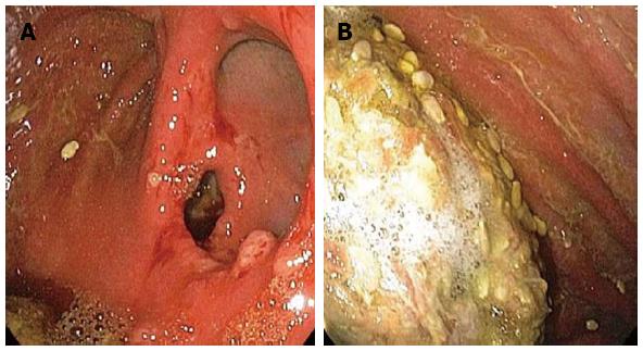
Figure 1 Esophagogastroduodenoscopy.
A: Polyps at the anastamosis; B: Gastric erythema and food bezoar.
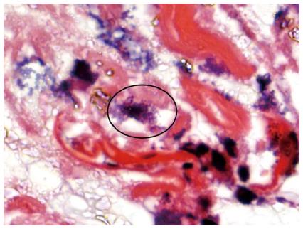
Figure 2 Characteristic 8-10 micron tetrads of Sarcina organisms (circled) were identified on endoscopic biopsy.
The background shows abundant bacterial overgrowth and debris from retained food. Separate fragments of ulcer bed were present (not pictured) (hematoxylin and eosin; original magnification × 1000 oil lens).
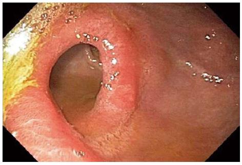
Figure 3 Repeat esophagogastroduodenoscopy showing improvement of gastric erythema.
-
Citation: Ratuapli SK, Lam-Himlin DM, Heigh RI.
Sarcina ventriculi of the stomach: A case report. World J Gastroenterol 2013; 19(14): 2282-2285 - URL: https://www.wjgnet.com/1007-9327/full/v19/i14/2282.htm
- DOI: https://dx.doi.org/10.3748/wjg.v19.i14.2282


