Copyright
©2012 Baishideng Publishing Group Co.
World J Gastroenterol. Feb 21, 2012; 18(7): 601-608
Published online Feb 21, 2012. doi: 10.3748/wjg.v18.i7.601
Published online Feb 21, 2012. doi: 10.3748/wjg.v18.i7.601
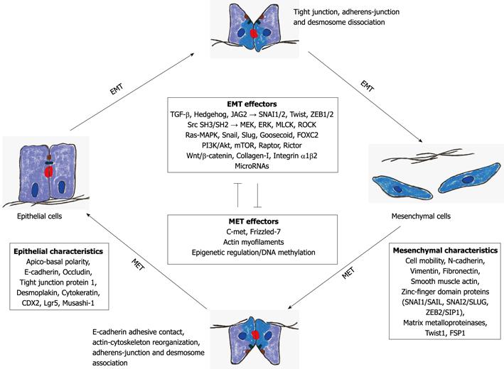
Figure 1 Schematic view of the effector pathways associated with epithelial-to-mesenchymal transition and mesenchymal-to-epithelial transition.
EMT: Epithelial-to-mesenchymal transition; MET: Mesenchymal-to-epithelial transition; TGF-β: Transforming growth factor-β; MAPK: Mitogen-activated protein kinase; ERK: Extracellular signal-regulated kinase; MEK: MAPK/ERK kinase; MLCK: Myosin light chain kinase; ROCK: Rho-dependent protein kinase.
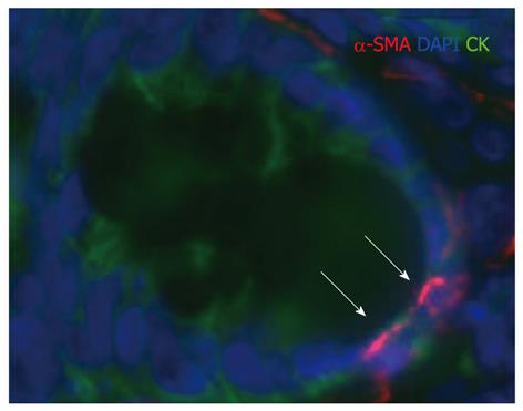
Figure 2 Chronic, fibrotizing phase of inflammatory bowel disease.
The arrowed cytokeratin (CK) positive epithelial cells in a colonic crypt show α-smooth muscle actin (α-SMA) positivity. The initiation of the epithelial-to-mesenchymal transition is possible. (α-SMA: Red; CK: Green; nuclear counter-staining: Blue; fluorescence immunohistochemistry, taken by virtual microscope). DAPI: 4′,6′-diamidino-2-phenylindole hydrochloride.
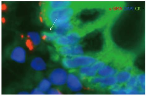
Figure 3 Regenerative phase of ulcerative colitis.
The arrowed α-SMA positive pericryptic cell shows cytokeratin (CK) expression. The presence of the mesenchymal-to-epithelial transition is possible. (α-SMA: Red; CK: Green; nuclear counter-staining: Blue; fluorescence immunohistochemistry, taken by virtual microscope). DAPI: 4′,6′-diamidino-2-phenylindole hydrochloride.
- Citation: Sipos F, Galamb O. Epithelial-to-mesenchymal and mesenchymal-to-epithelial transitions in the colon. World J Gastroenterol 2012; 18(7): 601-608
- URL: https://www.wjgnet.com/1007-9327/full/v18/i7/601.htm
- DOI: https://dx.doi.org/10.3748/wjg.v18.i7.601


