Copyright
©2012 Baishideng Publishing Group Co.
World J Gastroenterol. Dec 28, 2012; 18(48): 7394-7396
Published online Dec 28, 2012. doi: 10.3748/wjg.v18.i48.7394
Published online Dec 28, 2012. doi: 10.3748/wjg.v18.i48.7394
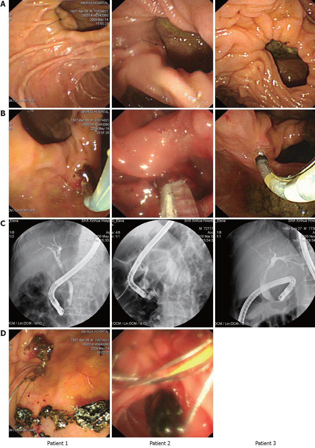
Figure 1 The duodenal diverticulum to be located intradiverticular papilla for biliary cannulation.
A: The papillary orifices can be indistinctly seen at the left side of the inner diverticular borders in patient 1 and patient 2 (arrow), but not seen at all in patient 3; B: Successful biliary cannulation was achieved after facing the intradiverticular papilla; C: Endoscopic retrograde cholangiography showing stones in the common bile duct in patient 1 and patient 2 and dilation of the common bile duct in patient 3; D: Extracted bile stones in the duodenal diverticulum in patient 1 and patient 2.
- Citation: Wang BC, Shi WB, Zhang WJ, Gu J, Tao YJ, Wang YQ, Wang XF. Entering the duodenal diverticulum: A method for cannulation of the intradiverticular papilla. World J Gastroenterol 2012; 18(48): 7394-7396
- URL: https://www.wjgnet.com/1007-9327/full/v18/i48/7394.htm
- DOI: https://dx.doi.org/10.3748/wjg.v18.i48.7394


