Copyright
©2012 Baishideng Publishing Group Co.
World J Gastroenterol. Dec 28, 2012; 18(48): 7201-7211
Published online Dec 28, 2012. doi: 10.3748/wjg.v18.i48.7201
Published online Dec 28, 2012. doi: 10.3748/wjg.v18.i48.7201
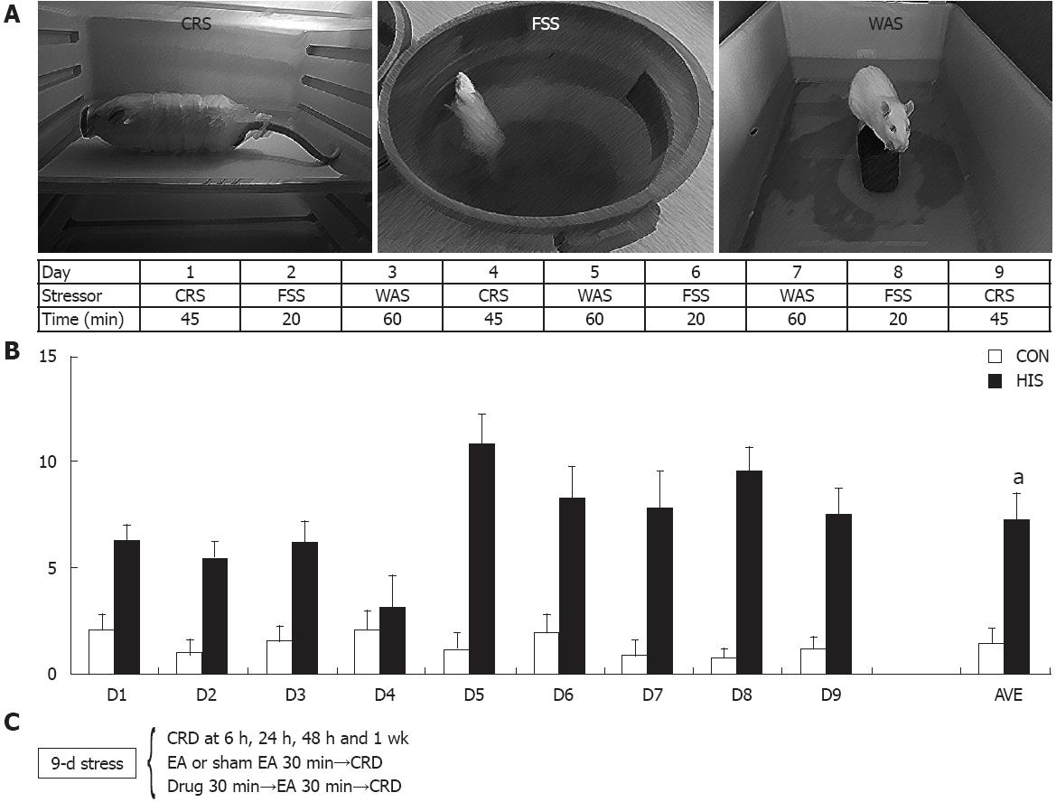
Figure 1 Heterotypic intermittent stress protocol and pellets count.
A: Nine-day heterotypic intermittent stress (HIS) protocol comprising three different randomly arranged stressors; B: Stress accelerated colonic transit (n = 10 for control, n = 8 for HIS), which was accompanied by an increase in visceral hypersensitivity (aP < 0.05). C: A schematic representation of the various treatments: stressors, colorectal distention (CRD) and drug treatment with time sequence. CON: Control; AVE: The mean number of pellets for all 9-d stress protocol or from respective control rats; CRS: Cold restrained stressor; FSS: Forced swimming stressor; WAS: Water avoidance stressor.
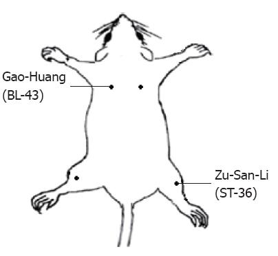
Figure 2 Schematic representation of electroacupuncture points.
ST-36 (Zu-San-Li) is thought to be relevant to gastrointestinal tract while Gao-Huang is not.
Figure 3 Heterotypic intermittent stress increased abdominal withdrawal reflex scores to colorectal distention.
A: Abdominal withdrawal reflex (AWR) scores were used as a function of distention pressure (20 mmHg, 40 mmHg, 60 mmHg and 80 mmHg). Heterotypic intermittent stress (HIS) significantly enhanced AWR scores measured at 6 h and 24 h after termination of last stressor when compared with baseline (Pre) under 20 mmHg, 60 mmHg and 80 mmHg distention pressure, while HIS significantly enhanced AWR scores measured at 6 h after termination of last stressor when compared with Pre group under 40 mmHg distention pressure [Tukey post hoc test following Friedman analysis of variance (ANOVA)]. Therefore, HIS can enhance visceral hypersensitivity in rats at 6 h and 24 h after termination of last stressor generally when compared with baseline (Pre). AWR scores returned to normal level 48 h after termination of last stressor (n = 8 rats for each group; aP < 0.05 vs Pre); B: HIS remarkably reduced distention threshold. Distention threshold was the minimal distention pressure to evoke abdominal movement. In agreement with AWR scores, distention threshold started to reduce at 6 h and maintained at a low level at 24 h and returned to normal level 48 h after termination of last stressor (Friedman ANOVA followed by Tukey post hoc test, n = 8 rats for each group; aP < 0.05 vs Pre).
Figure 4 Heterotypic intermittent stress enhanced electromyographic activities in response to colorectal distention.
Electromyographic (EMG) activities in the external oblique muscle in response to graded colorectal distention (CRD) were measured before stress and 6 h, 24 h, 48 h and 7 d after termination of heterotypic intermittent stress (HIS). The magnitude of EMG activity was expressed as area under curve (AUC). A: Examples of EMG activities recorded from baseline (Pre), 6 h, 24 h, 48 h and 7 d after termination of HIS in rats responding to distention pressures of 20 mmHg, 40 mmHg and 80 mmHg; B: Bar graph shows the changes in average of AUC before and after HIS protocol. There was no significant difference between EMGs of different time points at 20 mmHg. The magnitude of EMG activity of 6 h, 24 h and 48 h was significantly larger than that of Pre at distention pressures of 40 mmHg, 60 mmHg and 80 mmHg (Tukey post hoc test following two-way repeated measures analysis of variance, n = 6 rats for each group; aP < 0.05 vs Pre).
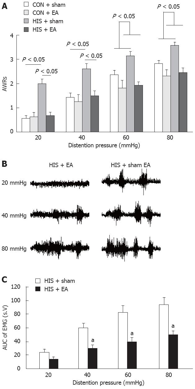
Figure 5 Electroacupuncture treatment attenuated the abdominal withdrawal reflex scores and electromyographic activities.
Electroacupuncture (EA) treatments were delivered for 30 min within 24 h after termination of last stressor. For sham EA group, the needle set was inserted into the ST-36 but no electrical stimulation was applied. A: The effects of stress exposure and EA treatment on abdominal withdrawal reflex (AWR) scores. Heterotypic intermittent stress (HIS) sham group showed a significant increase in AWR scores compared to control sham group under 20 mmHg and 40 mmHg distention pressures, while there was no significant difference in AWR scores between control and HIS groups after EA treatment. EA treatment at ST-36 point significantly decreased AWR scores in HIS rats, but had no effect on control rats under 20 mmHg and 40 mmHg pressures [Tukey post hoc test following two-way repeated measures analysis of variance (ANOVA)]. EA treatment significantly decreased AWR scores in both control and HIS rats under 60 mmHg and 80 mmHg pressures (two-way repeated measures ANOVA, n = 8 rats for each group; P < 0.05); B: Representative electromyographic (EMG) traces recorded immediately after termination of EA (left) or sham EA (right); C: Bar graph showing effects of EA treatment and sham EA on EMG recordings. The EMG was significantly decreased by EA treatment at pressure of 40 mmHg, 60 mmHg and 80 mmHg in stressed rats compared to sham EA groups (Tukey post hoc test following two-way repeated measures ANOVA, n = 6 rats for each group; aP < 0.05 vs HIS + sham group). CON: Control.
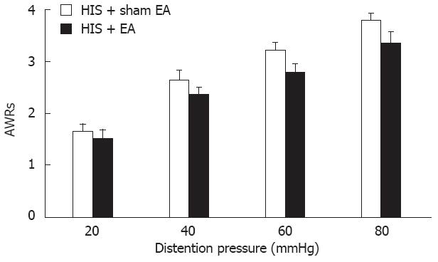
Figure 6 Effect of electroacupuncture at Gao-Huang.
Same electroacupuncture (EA) parameters as EA at ST-36 (Zu-San-Li) were used for BL-43 (Gao-Huang) treatment in heterotypic intermittent stress (HIS) rats. Although the abdominal withdrawal reflexes (AWRs) to colorectal distention heavily depend on the pressure level (two-way repeated measures analysis of variance, pressure effect, P < 0.001), EA treatment at BL-43 point on HIS rats showed no significant effect on pain perception; n = 7 rats for each group.
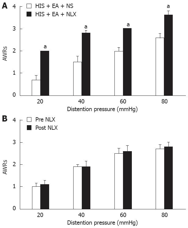
Figure 7 Reversal of electroacupuncture-mediated analgesic effect by naloxone.
A: The opioid receptor antagonist, naloxone (NLX) (0.1 mg/kg body weight, n = 5), or normal saline (NS) was administrated intraperitoneally 30 min before electroacupuncture (EA) application. Abdominal withdrawal reflex (AWR) scores were recorded immediately after EA termination in rats pretreated with NLX or NS. Both pressure and NLX injection significantly affected the AWRs to colorectal distention in rats [Friedman analysis of variance (ANOVA), P < 0.001 for pressure effect and for NLX injection effect]. Bar graph showed that NLX (0.1 mg/kg body weight) completely blocked the EA-induced analgesic effect when compared with NS treatment at all pressures (Mann-Whitney test following Friedman ANOVA). n = 5 rats for each group. aP < 0.05 vs NS-EA group; B: Same dose of NLX treatment did not produce any effect on AWR scores in control rats without EA treatment (Friedman ANOVA). AWRs were only significantly affected by different pressure levels (P < 0.001). n = 5 rats for each group. Control rats were not exposed to heterotypic intermittent stress (HIS).
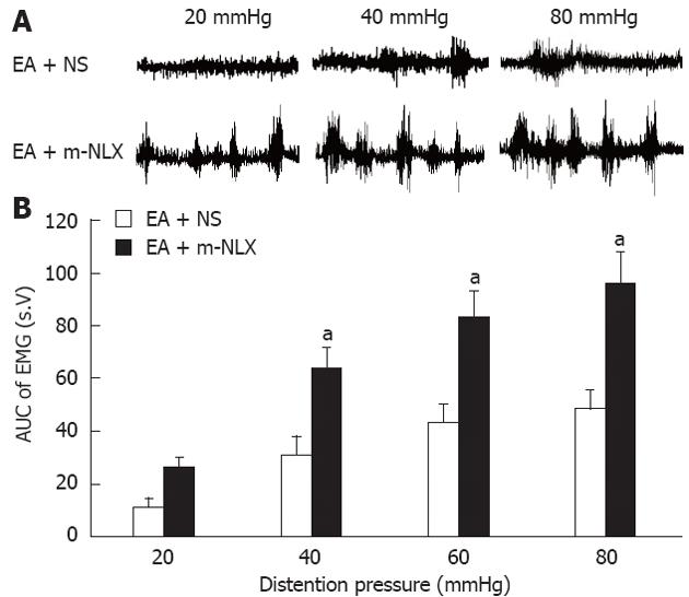
Figure 8 Reversal of electroacupuncture-mediated analgesic effect by naloxone methiodide.
The opioid receptor antagonist, naloxone methiodide (m-NLX), or normal saline (NS) was administrated intraperitoneally (5 mg/kg body weight) 30 min before electroacupuncture (EA) application. Electromyographic (EMG) activities were recorded immediately after EA from rats pretreated with m-NLX and NS. A: Examples of EA effects on EMG activities pretreated with NS (top) or m-NLX (bottom); B: Bar graph showing that m-NLX completely blocked the EA-induced analgesic effect. The magnitude of EMG activity was significantly increased by m-NLX treatment at pressure of 40 mmHg, 60 mmHg and 80 mmHg (Tukey post hoc test following two-way repeated measures analysis of variance, n = 6 rats for each group; aP < 0.05 vs EA + NS group).
- Citation: Zhou YY, Wanner NJ, Xiao Y, Shi XZ, Jiang XH, Gu JG, Xu GY. Electroacupuncture alleviates stress-induced visceral hypersensitivity through an opioid system in rats. World J Gastroenterol 2012; 18(48): 7201-7211
- URL: https://www.wjgnet.com/1007-9327/full/v18/i48/7201.htm
- DOI: https://dx.doi.org/10.3748/wjg.v18.i48.7201


