Copyright
©2012 Baishideng Publishing Group Co.
World J Gastroenterol. Dec 21, 2012; 18(47): 7127-7130
Published online Dec 21, 2012. doi: 10.3748/wjg.v18.i47.7127
Published online Dec 21, 2012. doi: 10.3748/wjg.v18.i47.7127
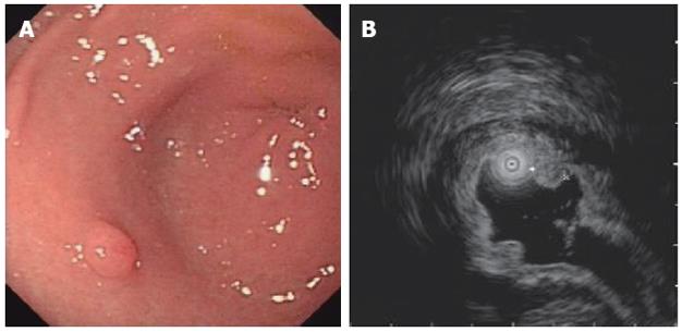
Figure 1 Subepithelial tumor in the stomach antrum.
A: Endoscopic examination showed a subepithelial tumor in the antrum; B: Endoscopic ultrasonography revealed that the tumor was localized in the mucosal layer.
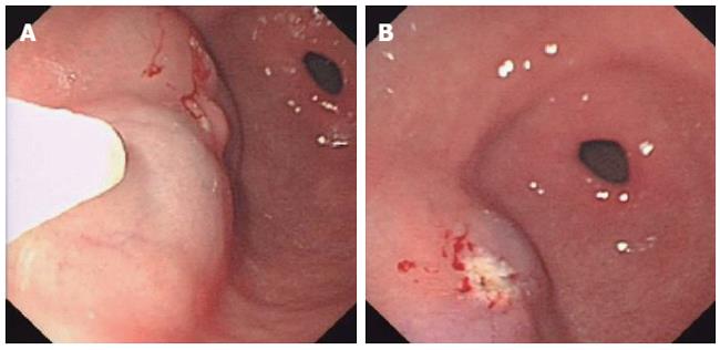
Figure 2 Endoscopic mucosal resection.
A: After injection of 15 mL saline, the whole nidus was lifted satisfactorily. However, an area of the uplifted mucosal layer was slightly blue; B: The lesion was successfully removed by high-frequency electrocoagulation, and a white wound was observed.
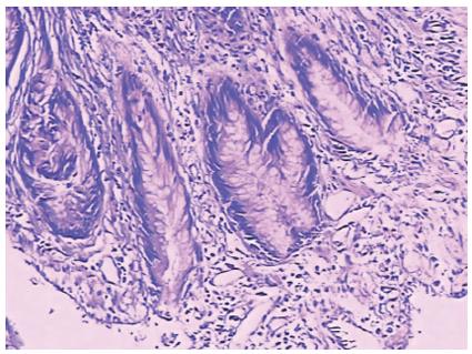
Figure 3 Pathologic examination revealed that the removed lesion from the gastric antrum was a tubular adenoma and Helicobacter pylori was not found.
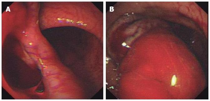
Figure 4 Emergency endoscopy.
A: A 4 cm × 5 cm diverticulum-like defect in anterior gastric antrum wall; B: A 4 cm × 8 cm intramural hematoma adjacent to the endoscopic submucosal dissection lesion and active bleeding.
- Citation: Sun P, Tan SY, Liao GH. Gastric intramural hematoma accompanied by severe epigastric pain and hematemesis after endoscopic mucosal resection. World J Gastroenterol 2012; 18(47): 7127-7130
- URL: https://www.wjgnet.com/1007-9327/full/v18/i47/7127.htm
- DOI: https://dx.doi.org/10.3748/wjg.v18.i47.7127


