Copyright
©2012 Baishideng Publishing Group Co.
World J Gastroenterol. Dec 21, 2012; 18(47): 6960-6966
Published online Dec 21, 2012. doi: 10.3748/wjg.v18.i47.6960
Published online Dec 21, 2012. doi: 10.3748/wjg.v18.i47.6960
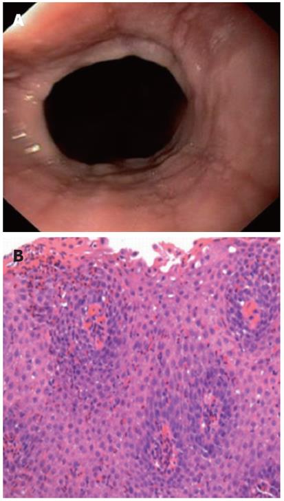
Figure 1 Endoscopic image and histological image of eosinophilic esophagitis.
A: Endoscopic image showing a lower esophageal Schatzki ring and linear furrowing of the esophageal mucosa, an endoscopic feature associated with eosinophilic esophagitis; B: Histological image of an esophageal biopsy, showing eosinophilic esophagitis with numerous intraepithelial eosinophils (> 50 eosinophils/high power field, hematoxylin and eosin, × 400).
- Citation: Müller M, Eckardt AJ, Fisseler-Eckhoff A, Haas S, Gockel I, Wehrmann T. Endoscopic findings in patients with Schatzki rings: Evidence for an association with eosinophilic esophagitis. World J Gastroenterol 2012; 18(47): 6960-6966
- URL: https://www.wjgnet.com/1007-9327/full/v18/i47/6960.htm
- DOI: https://dx.doi.org/10.3748/wjg.v18.i47.6960


