Copyright
©2012 Baishideng Publishing Group Co.
World J Gastroenterol. Nov 28, 2012; 18(44): 6510-6514
Published online Nov 28, 2012. doi: 10.3748/wjg.v18.i44.6510
Published online Nov 28, 2012. doi: 10.3748/wjg.v18.i44.6510
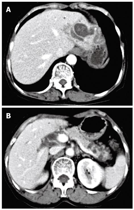
Figure 1 Computed tomography scan.
A: A cystic dilation of the left intrahepatic bile ducts accompanied with stones; B: Multiple cystic lesions in the pancreatic tail.
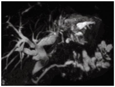
Figure 2 Magnetic resonance cholangiopancreatography reveals diffuse dilation of biliary tract with multiple filling defects in the left lateral lobe of the liver and segmental cystic dilation of pancreatic duct in the pancreatic tail.
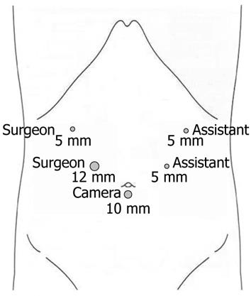
Figure 3 The location of trocars placement.
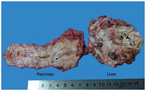
Figure 4 Resected specimens of intraductal papillary mucinous neoplasm-b (arrow head) and intraductal papillary mucinous neoplasm-p (arrow).
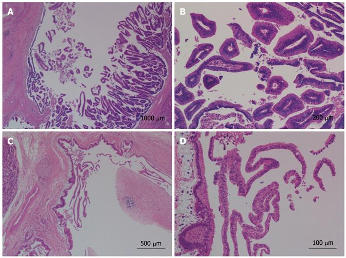
Figure 5 Histological features of intraductal papillary mucinous neoplasms by hematoxylin and eosin stain.
A: Liver, × 20; B: Liver, × 100; C: Pancreas, × 40; D: Pancreas, × 200.
- Citation: Xu XW, Li RH, Zhou W, Wang J, Zhang RC, Chen K, Mou YP. Laparoscopic resection of synchronous intraductal papillary mucinous neoplasms: A case report. World J Gastroenterol 2012; 18(44): 6510-6514
- URL: https://www.wjgnet.com/1007-9327/full/v18/i44/6510.htm
- DOI: https://dx.doi.org/10.3748/wjg.v18.i44.6510


