Copyright
©2012 Baishideng Publishing Group Co.
World J Gastroenterol. Nov 28, 2012; 18(44): 6510-6514
Published online Nov 28, 2012. doi: 10.3748/wjg.v18.i44.6510
Published online Nov 28, 2012. doi: 10.3748/wjg.v18.i44.6510
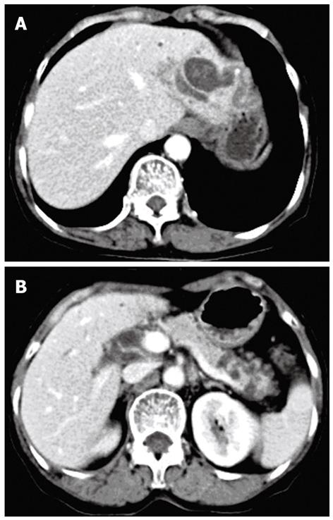
Figure 1 Computed tomography scan.
A: A cystic dilation of the left intrahepatic bile ducts accompanied with stones; B: Multiple cystic lesions in the pancreatic tail.
- Citation: Xu XW, Li RH, Zhou W, Wang J, Zhang RC, Chen K, Mou YP. Laparoscopic resection of synchronous intraductal papillary mucinous neoplasms: A case report. World J Gastroenterol 2012; 18(44): 6510-6514
- URL: https://www.wjgnet.com/1007-9327/full/v18/i44/6510.htm
- DOI: https://dx.doi.org/10.3748/wjg.v18.i44.6510


