Copyright
©2012 Baishideng Publishing Group Co.
World J Gastroenterol. Nov 21, 2012; 18(43): 6308-6314
Published online Nov 21, 2012. doi: 10.3748/wjg.v18.i43.6308
Published online Nov 21, 2012. doi: 10.3748/wjg.v18.i43.6308
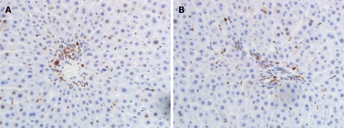
Figure 1 With prolongation of secondary biliary warm ischemia time, the number of proliferating-cell nuclear antigen-positive cholangiocytes was significantly reduced.
A: Proliferating-cell nuclear antigen (PCNA)-positive cholangiocytes in group II at 24 h after hepatic artery (HA) reperfusion; B: PCNA-positive cholangiocytes in group IV at 24 h after HA reperfusion. A and B, original magnification × 400.
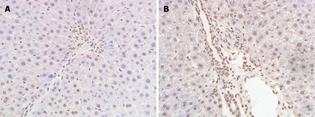
Figure 2 With prolongation of secondary biliary warm ischemia time, more biliary epithelial cells became apoptotic.
A: Biliary epithelial cell apoptosis in group II at 24 h after hepatic artery (HA) reperfusion; B: Biliary epithelial cell apoptosis in group IV at 24 h after HA reperfusion. A and B, original magnification × 400.
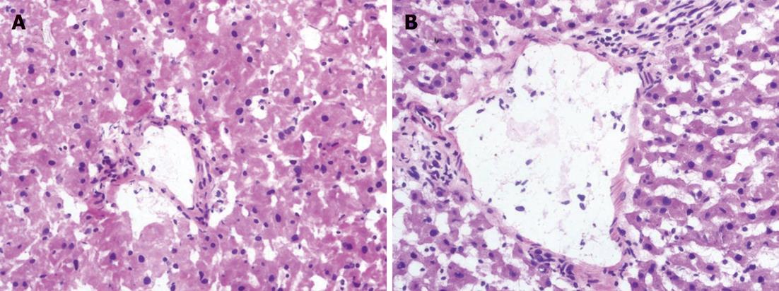
Figure 3 Histological examination of the liver at 24 h after hepatic arterial reperfusion.
A: Cholangiocyte injury can be found in group I; B: More marked injury occurs in group IV. A and B: Hematoxylin-eosin, original magnification × 400.
- Citation: Zhu XH, Pan JP, Wu YF, Ding YT. Effects of warm ischemia time on biliary injury in rat liver transplantation. World J Gastroenterol 2012; 18(43): 6308-6314
- URL: https://www.wjgnet.com/1007-9327/full/v18/i43/6308.htm
- DOI: https://dx.doi.org/10.3748/wjg.v18.i43.6308


