Copyright
©2012 Baishideng Publishing Group Co.
World J Gastroenterol. Oct 21, 2012; 18(39): 5551-5559
Published online Oct 21, 2012. doi: 10.3748/wjg.v18.i39.5551
Published online Oct 21, 2012. doi: 10.3748/wjg.v18.i39.5551
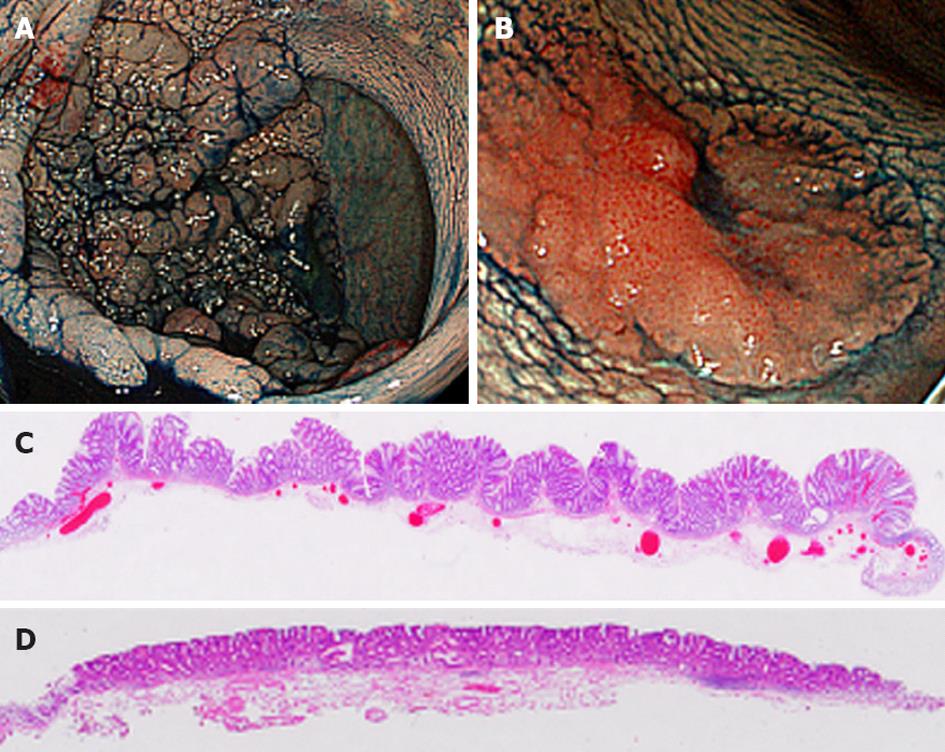
Figure 1 Macroscopic appearance (chromoscopic image) of laterally spreading tumors.
(A) Granular type (Gr-LST) and (B) flat- or non-granular type (NGr-LST). Whole mount view of cut sections of LSTs stained with hematoxylin and eosin: (C) A Gr-LST showing tubulovillous structures with nodular surfaces and (D) an NGr-LST consisting of small tubular glands with flat surfaces. LSTs: Laterally spreading tumors.
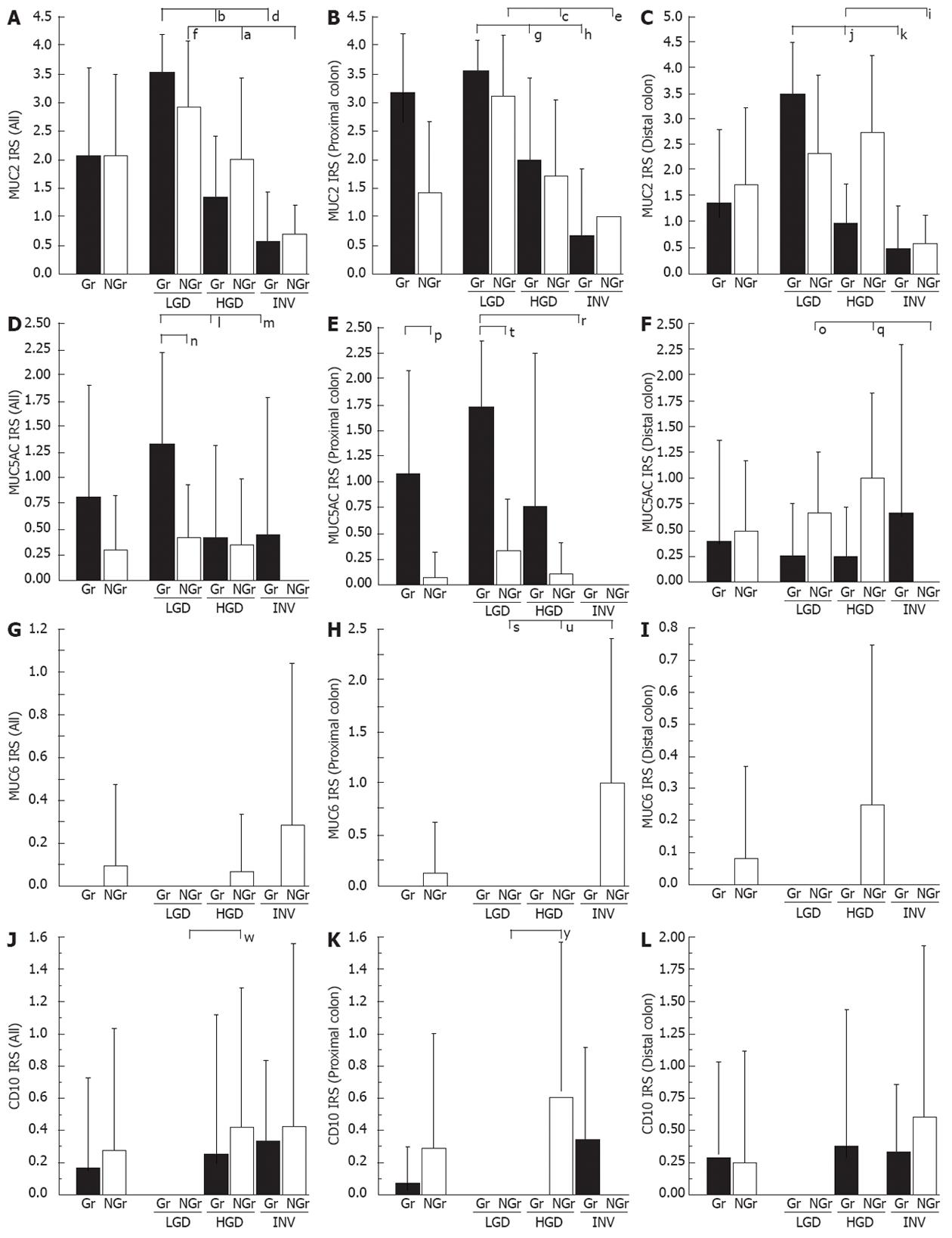
Figure 2 Immunoreactive scores for MUC2, MUC5AC, MUC6 and CD10 in all (A, D, G, J), proximal (B, E, H, K) and distal (C, F, I, L) laterally spreading tumors.
Gr, Gr-LST (black bar); NGr, NGr-LST (white bar). IRS: Immunoreactive score; LGD: Adenoma with low grade dysplasia; HGD: Adenoma with high grade dysplasia; INV: Adenocarcinoma invading into submucosa; LSTs: laterally spreading tumors; Gr: Granular; NGr: Non-granular. Data are mean ± SD; a, c, e, g, i, k, m, o, q, s, u, w, yP < 0.05, MUC2: all NGr-HGD vs NGr-INV; proximal colon NGr-LGD vs NGr-HGD or NGr-INV; Gr-LGD vs Gr-HGD; distal colon Gr-LGD vs Gr-INV; NGr-HGD vs NGr-INV; MUC5AC: all Gr-LGD vs Gr-INV; distal colon NGr-INV vs NGr-HGD or NGr-LGD; MUC6: proximal colon NGr-INV vs NGr-LGD or NGr-HGD; CD10: all NGr-LGD vs NGr-HGD; proximal colon NGr-LGD vs NGr-HGD; b, d, f, h, j, l, n, p, r, tP < 0.01, MUC2: all Gr-LGD vs Gr-HGD or Gr-INV; NGr-LGD vs NGr-INV; proximal colon Gr-LGD vs Gr-INV; distal colon Gr-LGD vs Gr-HGD; MUC5AC: all Gr-LGD vs NGr-LGD; Gr-LGD vs Gr-HGD; proximal colon Gr-type vs NGr-type; Gr-LGD vs NGr-LGD; Gr-LGD vs Gr-INV.
Figure 3 Immunoreactive scores for p53 (A) and nuclear β-catenin (B), and labeling indices for Ki-67 in the lower zone (C) in all laterally spreading tumors.
Gr, Gr-LST (black bar); NGr, NGr-LST (white bar); LSTs: laterally spreading tumors. Data are mean ± SD; a, c, eP < 0.05, p53: Gr-HGD vs Gr-INV; nuclear β-catenin: Gr-type vs NGr-type; Ki-67, Gr-type vs NGr-type; b, d, f, h, j, l, nP < 0.01, p53: NGr-LGD vs NGr-INV; Gr-LGD vs Gr-HGD or Gr-INV; nuclear β-catenin: Gr-LGD vs Gr-HGD or Gr-INV: Gr-LGD vs NGr-LGD; Ki-67, Gr-LGD vs NGr-INV. IRSs: Immunoreactive scores; LGD: Adenoma with low grade dysplasia; HGD: Adenoma with high grade dysplasia; INV: Adenocarcinoma invading into submucosa; Gr: Granular; NGr: Non-granular; LI: Labeling indice.
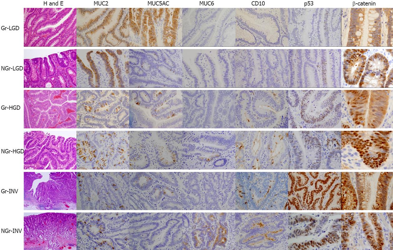
Figure 4 Histology and immunohistochemistry of laterally spreading tumors.
Immunoreactive scores (IRSs) for Gr-LGD: MUC2, 4 points; MUC5AC, 2 points; MUC6, 0 points; CD10, 0 points; p53, 0 points; nuclear β-catenin, 0 points. IRSs for NGr-LGD: MUC2, 3 points; MUC5AC, 1 points; MUC6, 0 points, CD10, 0 points; p53, 0 points; nuclear β-catenin, 1 points. IRSs for Gr-HGD: MUC2, 1 points; MUC5AC, 1 points; MUC6, 0 points, CD10, 1 points; p53, 1 points; nuclear β-catenin, 1 points. IRSs for NGr-HGD: MUC2, 2 points; MUC5AC, 1 points; MUC6, 1 points, CD10, 1 points; p53, 1 points; nuclear β-catenin, 2 points. IRSs for Gr-INV: MUC2, 1 points; MUC5AC, 1 points; MUC6, 0 points, CD10, 1 points; p53, 4 points; nuclear β-catenin, 1 points. IRSs for NGr-INV: MUC2, 1 points; MUC5AC, 0 points; MUC6, 2 points, CD10, 2 points; p53, 2 points; nuclear β-catenin, 2 points (H and E, ×80; immunoperoxidase: MUC2, MUC5AC and MUC6, ×100; CD10 and p53, ×120; β-catenin, ×300); LGD: Adenoma with low grade dysplasia; HGD: Adenoma with high grade dysplasia; INV: Adenocarcinoma invading into submucosa; Gr: Granular; NGr: Non-granular.
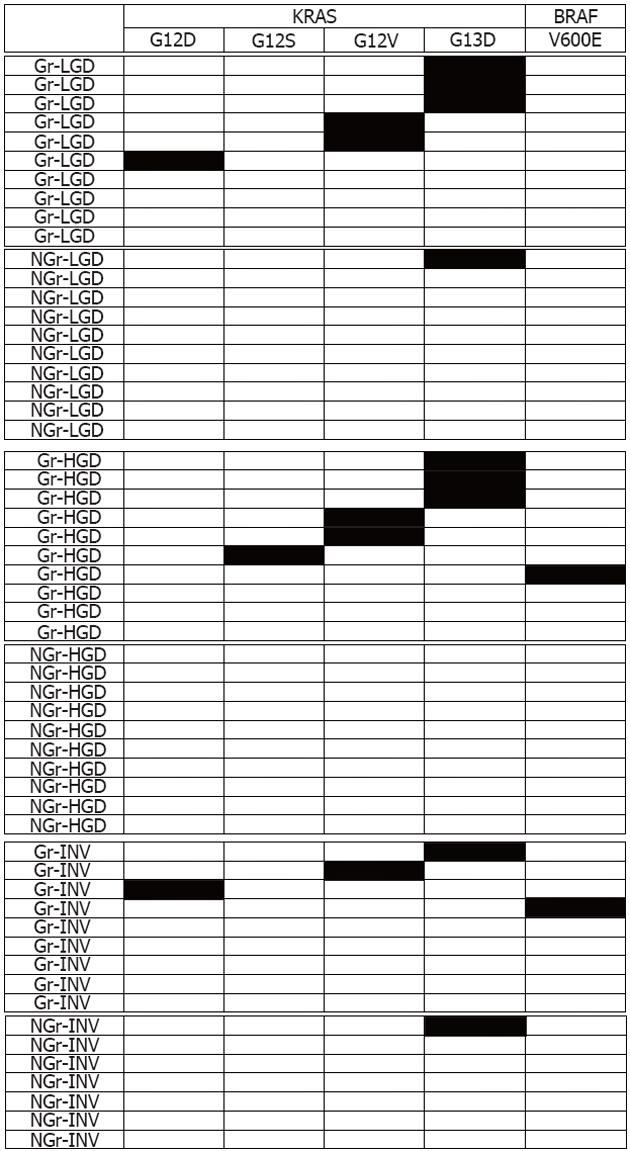
Figure 5 v-Ki-ras2 Kirsten rat sarcoma viral oncogene homolog and v-raf murine sarcoma viral oncogene homologue B1 mutational patterns in laterally spreading tumors.
D: Aspartic acid; E: Glutamic acid; G: Glycine; S: Serine; V: Valine; Black box: Mutated case; White box: Non-mutated case; LGD: Adenoma with low grade dysplasia; HGD: Adenoma with high grade dysplasia; INV: Adenocarcinoma invading into submucosa; Gr: Granular; NGr: Non-granular; KRAS: v-Ki-ras2 Kirsten rat sarcoma viral oncogene homolog; BRAF: v-raf murine sarcoma viral oncogene homologue B1.
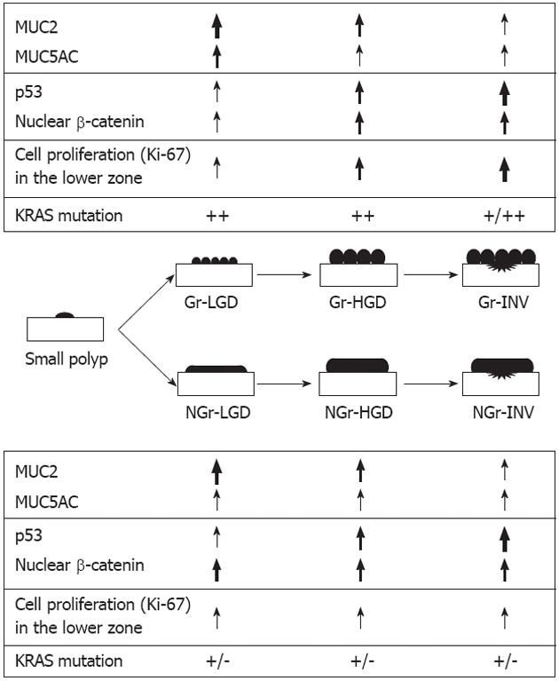
Figure 6 Alterations of expression of mucin core protein, p53 and β-catenin, cell proliferation and v-Ki-ras2 Kirsten rat sarcoma viral oncogene homolog mutations in malignant transformation of laterally spreading tumors.
Large arrow: Marked upregulation; Medium arrow: Moderate upregulation; Small arrow: Mild upregulation; ++: Frequently mutated; +: Infrequently mutated; -: Not mutated; LGD: Adenoma with low grade dysplasia; HGD: Adenoma with high grade dysplasia; INV: Adenocarcinoma invading into submucosa; KRAS: v-Ki-ras2 Kirsten rat sarcoma viral oncogene homolog; Gr: Granular; NGr: Non-granular.
-
Citation: Nakae K, Mitomi H, Saito T, Takahashi M, Morimoto T, Hidaka Y, Sakamoto N, Yao T, Watanabe S. MUC5AC/β-catenin expression and
KRAS gene alteration in laterally spreading colorectal tumors. World J Gastroenterol 2012; 18(39): 5551-5559 - URL: https://www.wjgnet.com/1007-9327/full/v18/i39/5551.htm
- DOI: https://dx.doi.org/10.3748/wjg.v18.i39.5551


