Copyright
©2012 Baishideng Publishing Group Co.
World J Gastroenterol. Sep 28, 2012; 18(36): 4985-4993
Published online Sep 28, 2012. doi: 10.3748/wjg.v18.i36.4985
Published online Sep 28, 2012. doi: 10.3748/wjg.v18.i36.4985
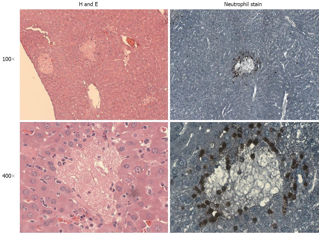
Figure 1 Sample liver sections from C57Bl/6 mice illustrating neutrophil accumulation after bile duct ligation.
C57BL/6 mice were subjected to bile duct ligation and sacrificed 24 h later. Hematoxylin and eosin (H and E) stained sections showing focal necrosis at 100× and 400× magnification. Sections stained with an anti-neutrophil antibody. Neutrophils are present largely around areas of focal necrosis in this model.
- Citation: Woolbright BL, Jaeschke H. Novel insight into mechanisms of cholestatic liver injury. World J Gastroenterol 2012; 18(36): 4985-4993
- URL: https://www.wjgnet.com/1007-9327/full/v18/i36/4985.htm
- DOI: https://dx.doi.org/10.3748/wjg.v18.i36.4985


