Copyright
©2012 Baishideng Publishing Group Co.
World J Gastroenterol. Sep 21, 2012; 18(35): 4866-4874
Published online Sep 21, 2012. doi: 10.3748/wjg.v18.i35.4866
Published online Sep 21, 2012. doi: 10.3748/wjg.v18.i35.4866
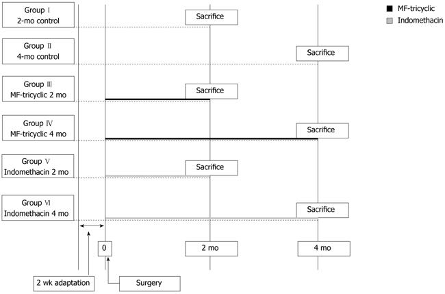
Figure 1 Scheme of the study design.
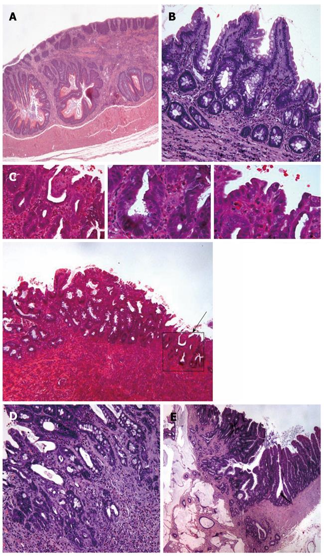
Figure 2 Pathological findings in the esophagus of rats with esophagojejunostomy.
A: Hyperplasia of squamous epithelium in a control rat esophagus; B: Hematoxylin and eosin (HE) staining (× 100) showing an area of intestinal metaplasia without dysplasia. The top picture shows in more detail a gland (× 400); C: Indomethacin-treated rat. HE staining (× 40) of an area of intestinal metaplasia showing a focus with low-grade dysplasia (black arrow). The top pictures show in more detail the area with low grade dysplasia (× 200; × 400). Glands conserve their architecture. Nuclear pseudostratification can be seen, nuclear size is also increased but nuclei do not reach the lumen; D: HE staining of an area of intestinal metaplasia showing high grade dysplasia. Magnification × 100; the top picture illustrates at higher magnification (× 400), some glands with high grade dysplasia, characterized by loss of nuclear polarity and atypia; E: Photomicrograph shows an adenocarcinoma developed inside the intestinal metaplasia of the esophagus near the jejunum epithelium in a rat that had undergone an esophagojejunostomy. HE staining shows that the esophageal adenocarcinoma consists of dilated, cystic glands with abundant mucin secretion and epithelial dysplasia. (Magnification × 40; the top picture shows the same fields at × 100 magnification).
Figure 3 Macroscopic findings.
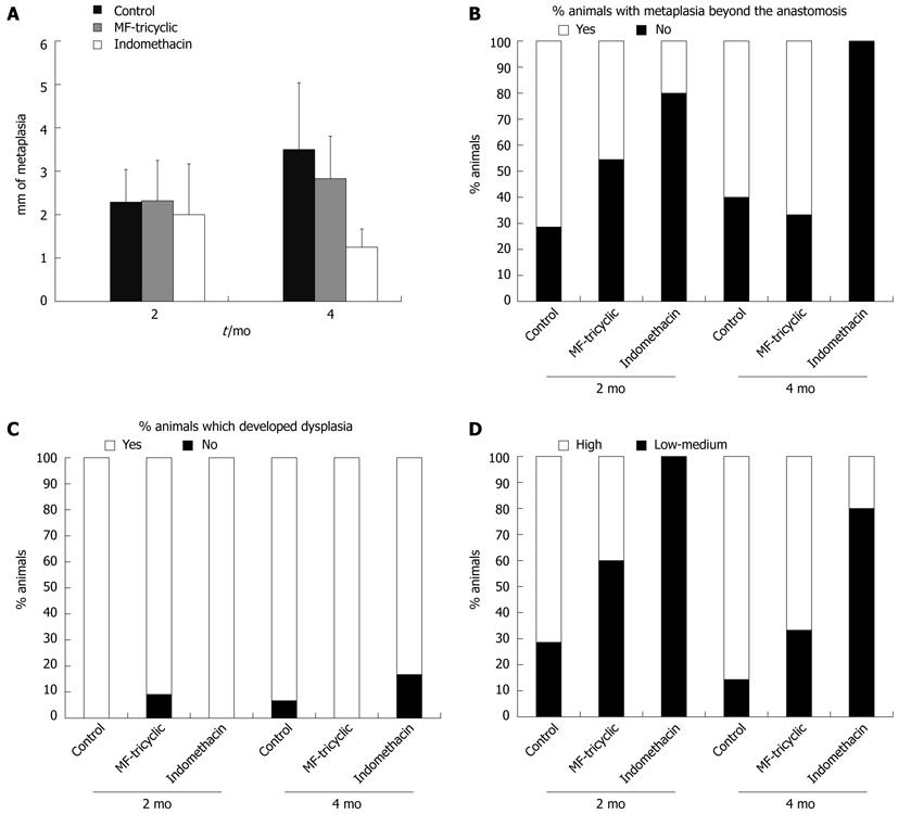
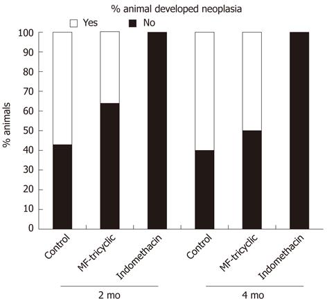
A: Effect of treatment on metaplasia in continuity length. A significant decrease was observed in indomethacin-treated groups compared to control groups (P = 0.007); B: Treatment effect on the percentage of animals that developed metaplasia beyond the anastomosis. Percentage of animals that developed metaplasia beyond the anastomosis was significantly higher in both control and MF-tricyclic groups (P = 0.009 and P = 0.036, respectively); C: Percentage of animals that developed dysplasia; D: Because most animals developed dysplasia, low-to-medium grade vs high-grade dysplasia was analyzed. Percentage of rats that developed high-grade dysplasia was significantly lower in indomethacin groups compared to control groups (P = 0.002). When we analyzed the effect of treatment, we observed lower incidence of neoplasia in the indomethacin group (P = 0.000). MF-tricyclic: 3-(3, 4-difluorophenyl)-4-(4-(methylsulfonyl)phenyl)-2-(5H)-furanone.
Figure 4 Percentage of animals that developed neoplasia.
- Citation: Esquivias P, Morandeira A, Escartín A, Cebrián C, Santander S, Esteva F, García-González MA, Ortego J, Lanas A, Piazuelo E. Indomethacin but not a selective cyclooxygenase-2 inhibitor inhibits esophageal adenocarcinogenesis in rats. World J Gastroenterol 2012; 18(35): 4866-4874
- URL: https://www.wjgnet.com/1007-9327/full/v18/i35/4866.htm
- DOI: https://dx.doi.org/10.3748/wjg.v18.i35.4866


