Copyright
©2012 Baishideng Publishing Group Co.
World J Gastroenterol. Sep 14, 2012; 18(34): 4794-4797
Published online Sep 14, 2012. doi: 10.3748/wjg.v18.i34.4794
Published online Sep 14, 2012. doi: 10.3748/wjg.v18.i34.4794
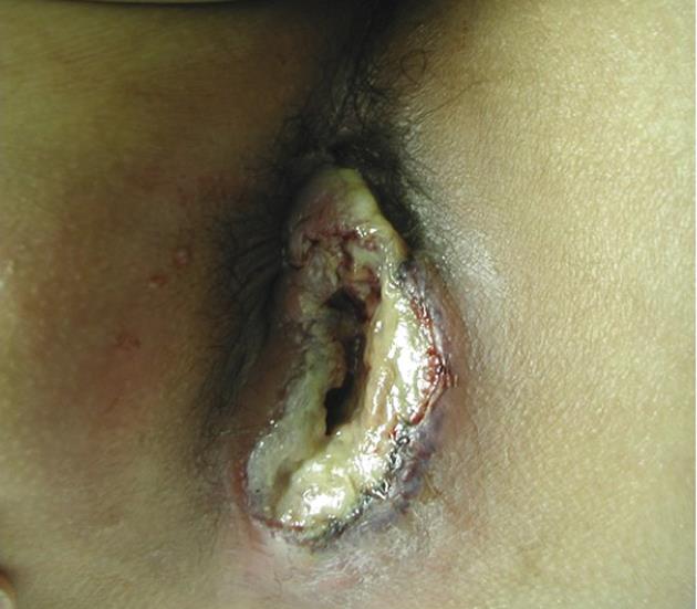
Figure 1 A giant ulcer covered with necrotic tissue is identified in the perianal region.
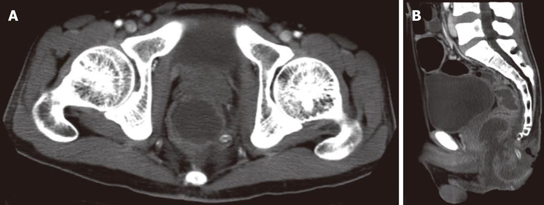
Figure 2 Abdominal computed tomography revealing an abscess formation of the perianal region.
A: The axial view shows an abscess of the perianal region due to necrosis; B: The sagittal view shows an abscess formation extending from the perianal region to the lower rectum.
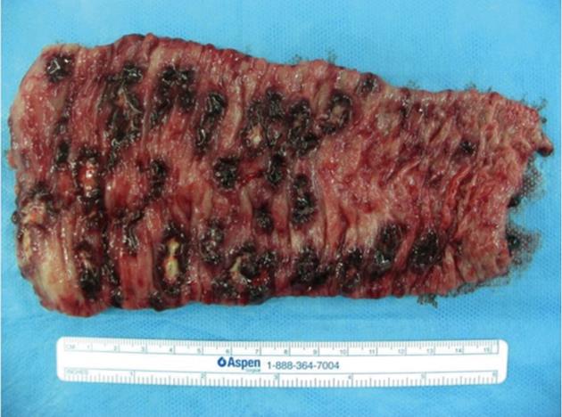
Figure 3 Macroscopic findings of the resected colon following end-sigmoidostomy show numerous deep ulcerations covered with yellow necrotic plaques.
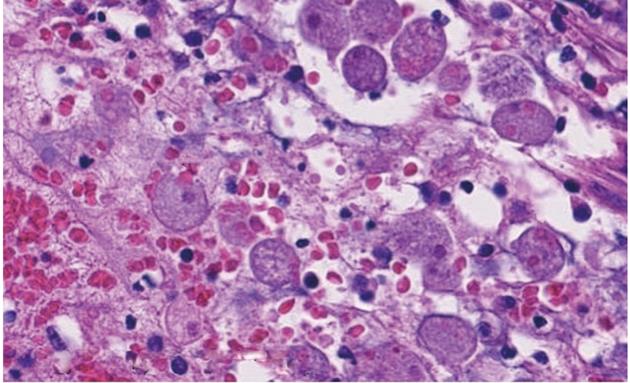
Figure 4 Histopathological findings of the surgical specimen show trophozoite amoebae that have ingested red blood cells (arrows) (HE staining).
- Citation: Torigoe T, Nakayama Y, Yamaguchi K. Development of perianal ulcer as a result of acute fulminant amoebic colitis. World J Gastroenterol 2012; 18(34): 4794-4797
- URL: https://www.wjgnet.com/1007-9327/full/v18/i34/4794.htm
- DOI: https://dx.doi.org/10.3748/wjg.v18.i34.4794


