Copyright
©2012 Baishideng Publishing Group Co.
World J Gastroenterol. Sep 14, 2012; 18(34): 4639-4650
Published online Sep 14, 2012. doi: 10.3748/wjg.v18.i34.4639
Published online Sep 14, 2012. doi: 10.3748/wjg.v18.i34.4639
Figure 1 Professor Pablo Luis Mirizzi (1893-1964).
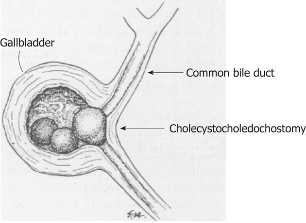
Figure 2 Albert Behrend described this case that corresponded to Mirizzi syndrome type IV according to the Csendes classification.
The gallstone surrounded by an inflammatory process was clearly dilating the cystic duct fusing the gallbladder to the bile duct (original drawing from Annals of Surgery 1950; 132, page 300)[20].
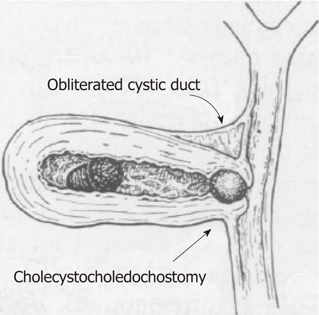
Figure 3 Another case described by Behrend.
The cystic duct has become obliterated due to a chronic inflammatory process and the gallstones have eroded the gallbladder wall and formed a cholecystobiliary fistula corresponding to Mirizzi syndrome type II according to the Csendes classification (original drawing from Annals of Surgery 1950; 132, page 300)[20].
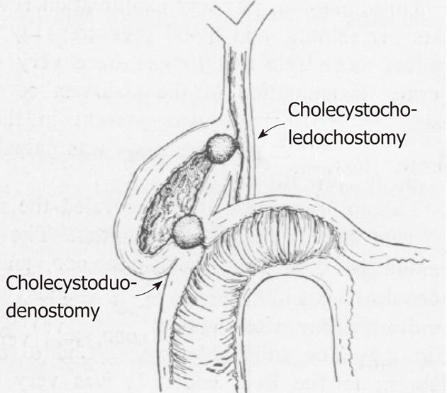
Figure 4 This illustration by Behrend clearly depicts Mirizzi syndrome type V according to Csendes.
The continuous inflammatory process affecting the gallbladder has obliterated the cystic duct, and the gallstones compressed inside the gallbladder, have produced pressure ulcers over the gallbladder walls eroding into the bile duct and the duodenum (original drawing from Annals of Surgery 1950; 132, page 300)[20].
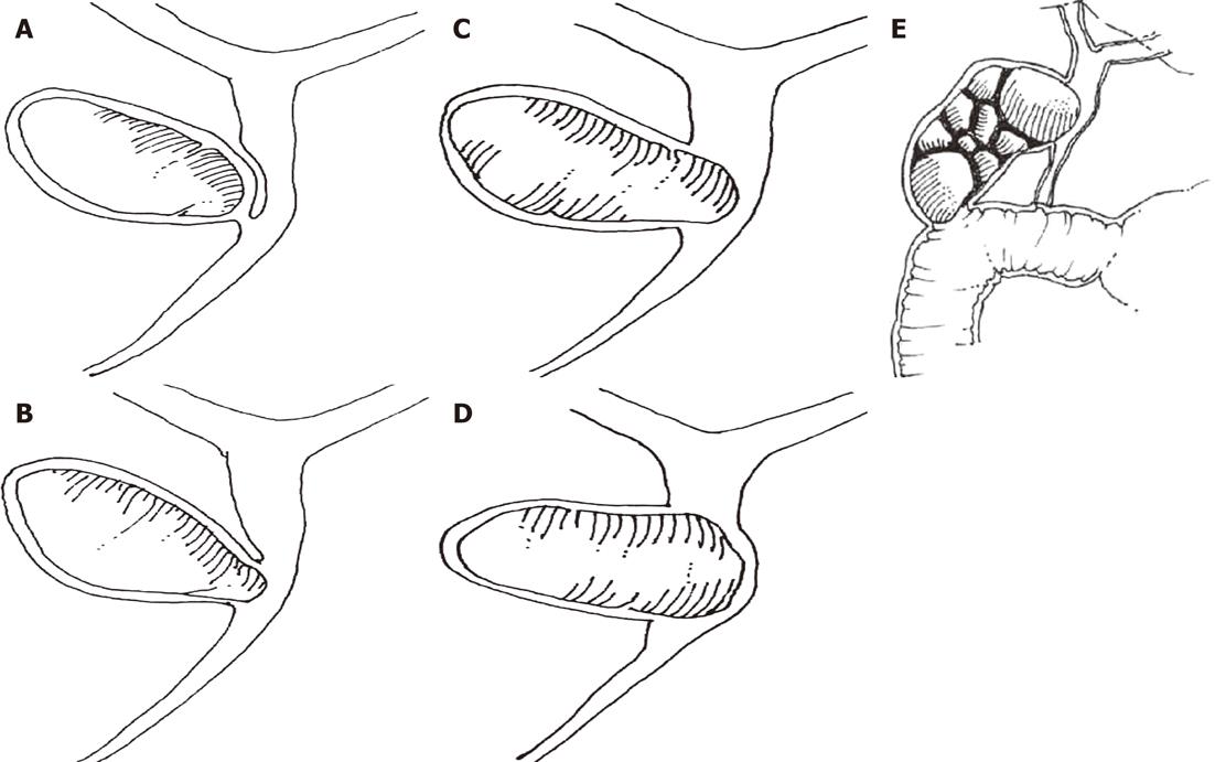
Figure 5 Mirizzi syndrome type I to type V.
A: Mirizzi type I is the extrinsic compression of the common bile duct by an impacted gallstone; B: Mirizzi type II consists of a cholecystobiliary fistula involving one third of the circumference of the bile duct; C: In Mirizzi type III, the cholecystobiliary fistula compromises up to two-thirds of the circumference of the bile duct; D: In Mirizzi type IV, the cholecystobiliary fistula has destroyed the bile duct wall, and comprises the whole circumference of the bile duct; E: Mirizzi type V corresponds to any type of Mirizzi associated with a bilioenteric fistula with or without gallstone ileus (original drawings from World Journal of Surgery 2008; 32, pages 2239 to 2241)[4].
- Citation: Beltrán MA. Mirizzi syndrome: History, current knowledge and proposal of a simplified classification. World J Gastroenterol 2012; 18(34): 4639-4650
- URL: https://www.wjgnet.com/1007-9327/full/v18/i34/4639.htm
- DOI: https://dx.doi.org/10.3748/wjg.v18.i34.4639


