Copyright
©2012 Baishideng Publishing Group Co.
World J Gastroenterol. Aug 7, 2012; 18(29): 3883-3888
Published online Aug 7, 2012. doi: 10.3748/wjg.v18.i29.3883
Published online Aug 7, 2012. doi: 10.3748/wjg.v18.i29.3883
Figure 1 Comparison of endoscopic ultrasound-guided fine needle aspiration with International Consensus Diagnostic Criteria.
EUS-FNA: Endoscopic ultra-sound-guided fine needle aspiration; LPSP: Lymphoplasmacytic sclerosing pancreatitis; IDCP: Idiopathic duct-centric pancreatitis; OOI: Other organ involvement; AIP: Autoimmune pancreatitis; NOS: Not otherwise specified.
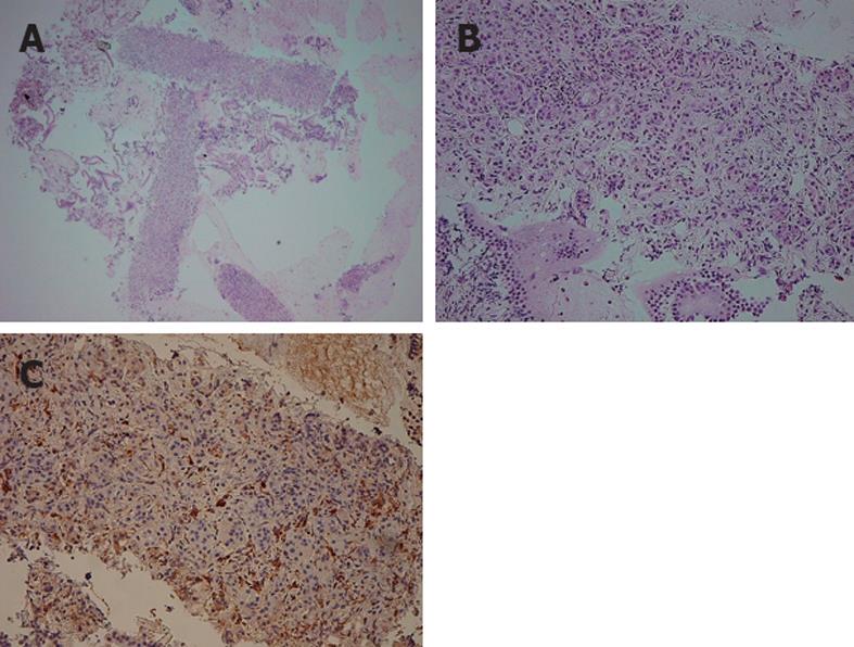
Figure 2 Endoscopic ultrasound-guided fine needle aspiration specimen of "definitive type 1 autoimmune pancreatitis”.
A, B: Hematoxylin and eosin staining of a resected pancreas specimen obtained by endoscopic ultra-sound-guided fine needle aspiration shows replacement of the acinar structure by lymphoplasmacytic infiltration and fibrosis; C: Numerous plasma cells show positive immunoreactivity for immunoglobulin G4.
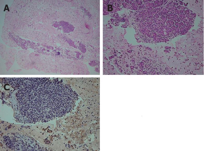
Figure 3 Endoscopic ultrasound-guided fine needle aspiration specimen of "probable type 2 autoimmune pancreatitis”.
A, B: Hematoxylin and eosin staining of a resected pancreas specimen obtained by endoscopic ultra-sound-guided fine needle aspiration shows the infiltration of neutrophils in addition to lymphocyte infiltration and fibrosis; C: Immunostaining for immunoglobulin G4 is negative.
- Citation: Ishikawa T, Itoh A, Kawashima H, Ohno E, Matsubara H, Itoh Y, Nakamura Y, Hiramatsu T, Nakamura M, Miyahara R, Ohmiya N, Goto H, Hirooka Y. Endoscopic ultrasound-guided fine needle aspiration in the differentiation of type 1 and type 2 autoimmune pancreatitis. World J Gastroenterol 2012; 18(29): 3883-3888
- URL: https://www.wjgnet.com/1007-9327/full/v18/i29/3883.htm
- DOI: https://dx.doi.org/10.3748/wjg.v18.i29.3883


