Copyright
©2012 Baishideng Publishing Group Co.
World J Gastroenterol. Jul 28, 2012; 18(28): 3721-3726
Published online Jul 28, 2012. doi: 10.3748/wjg.v18.i28.3721
Published online Jul 28, 2012. doi: 10.3748/wjg.v18.i28.3721
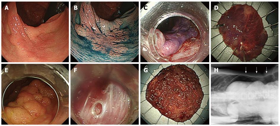
Figure 1 Case without complications and with perforation.
A: Tumor located in the ascending colon; B: Image of dye spraying (indigo carmine). The macroscopic type is 0-IIa (LST-NG). The size is 35 mm; C: Treatment by endoscopic submucosal dissection (ESD); D: En-bloc resection was performed; E: Tumor located in the cecum. The macroscopic type is 0-IIa (LST-G). The size is 65 mm; F: Perforation occurred during ESD. It was closed by endoscopic clipping; G: En-bloc resection was performed by ESD; H: Prominent free air was observed in the abdominal cavity with the patient lying on the left side. The free air is indicated in the Figure by an arrow. LST-NG: Laterally spreading tumors-non-granular; LST-G: Laterally spreading tumors-granular.
- Citation: Aoki T, Nakajima T, Saito Y, Matsuda T, Sakamoto T, Itoi T, Khiyar Y, Moriyasu F. Assessment of the validity of the clinical pathway for colon endoscopic submucosal dissection. World J Gastroenterol 2012; 18(28): 3721-3726
- URL: https://www.wjgnet.com/1007-9327/full/v18/i28/3721.htm
- DOI: https://dx.doi.org/10.3748/wjg.v18.i28.3721


