Copyright
©2012 Baishideng Publishing Group Co.
World J Gastroenterol. Jul 28, 2012; 18(28): 3673-3680
Published online Jul 28, 2012. doi: 10.3748/wjg.v18.i28.3673
Published online Jul 28, 2012. doi: 10.3748/wjg.v18.i28.3673
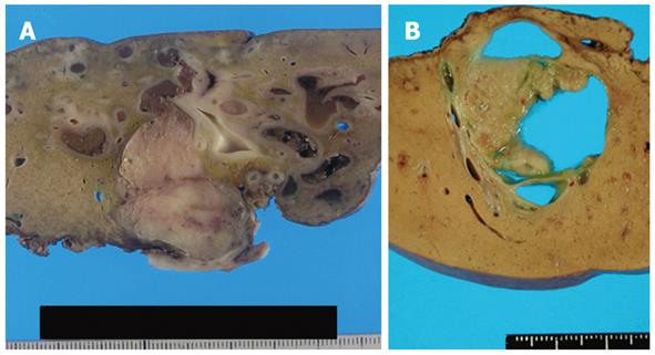
Figure 1 Gross findings.
A: Duct-ectatic type. Tumor filled the dilated intrahepatic bile duct. Surrounding bile duct was also dilated; B: Cystic type. Cystic dilatation of intrahepatic bile duct. Papillary tumor was found in the dilated intrahepatic bile duct. Significant retention of mucin was observed.
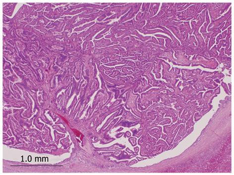
Figure 2 Histopathology of intraductal neoplasm of the intrahepatic bile duct.
Tumor shows papillary proliferation within the dilated bile duct (hematoxylin and eosin stain, × 20).
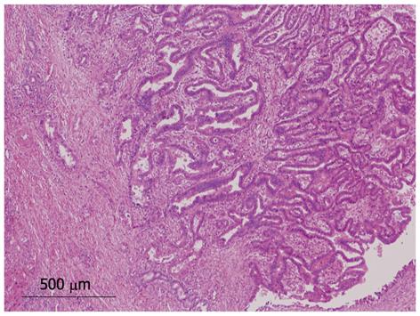
Figure 3 Microinvasion of tumor.
Tumor cells infiltrating into the bile duct wall (hematoxylin and eosin stain, × 40).
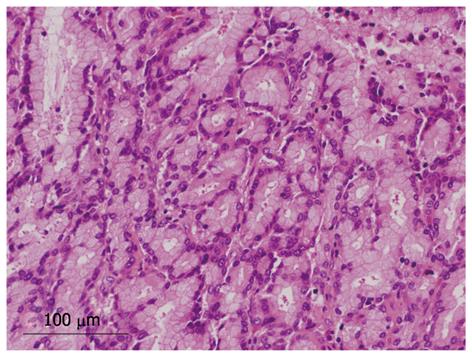
Figure 4 Tubular structure within intraductal neoplasm of the intrahepatic bile duct.
Cells with intracellular mucin and mild atypia forming a pyloric gland-like structure (hematoxylin and eosin stain, × 200).
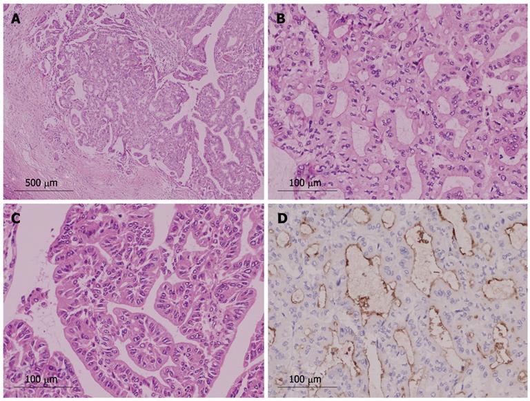
Figure 5 Histology of intraductal neoplasm of the intrahepatic bile duct with papillary and tubular structure (case 1).
A: Histological structure was mainly tubular, but papillary structure was also present [hematoxylin and eosin (HE) stain, × 40]; B: Tubular structure (HE stain, × 200); C: Papillary structure (HE stain, × 200); D: Tumor cells were positive for mucin (MUC)1 immunohistochemistry (MUC1 stain, × 200).
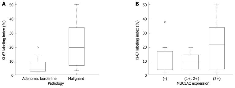
Figure 6 Box plot for Ki-67 labeling index by histological degree of malignancy and MUC5AC expression.
A: The Ki-67 labeling index (LI) was significantly higher in the malignant group than in the benign/borderline group; B: There was an association with borderline significance between MUC5AC expression and Ki-67 LI (P = 0.0622). MUC: Mucin.
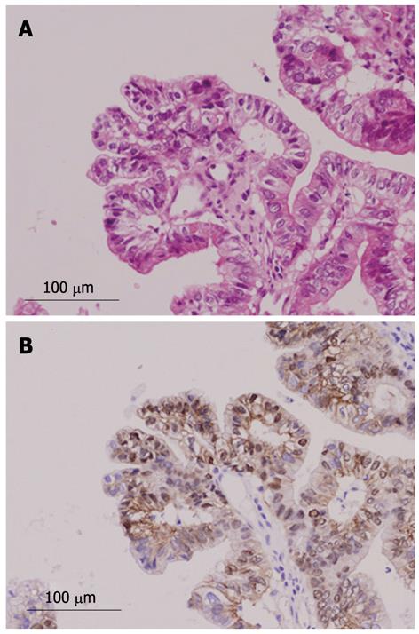
Figure 7 Nuclear expression of β-catenin in pancreaticobiliary type.
A: Hematoxylin and eosin stain, × 200; B: β-catenin stain, × 200.
- Citation: Naito Y, Kusano H, Nakashima O, Sadashima E, Hattori S, Taira T, Kawahara A, Okabe Y, Shimamatsu K, Taguchi J, Momosaki S, Irie K, Yamaguchi R, Yokomizo H, Nagamine M, Fukuda S, Sugiyama S, Nishida N, Higaki K, Yoshitomi M, Yasunaga M, Okuda K, Kinoshita H, Nakayama M, Yasumoto M, Akiba J, Kage M, Yano H. Intraductal neoplasm of the intrahepatic bile duct: Clinicopathological study of 24 cases. World J Gastroenterol 2012; 18(28): 3673-3680
- URL: https://www.wjgnet.com/1007-9327/full/v18/i28/3673.htm
- DOI: https://dx.doi.org/10.3748/wjg.v18.i28.3673


