Copyright
©2012 Baishideng Publishing Group Co.
World J Gastroenterol. Jan 14, 2012; 18(2): 126-135
Published online Jan 14, 2012. doi: 10.3748/wjg.v18.i2.126
Published online Jan 14, 2012. doi: 10.3748/wjg.v18.i2.126
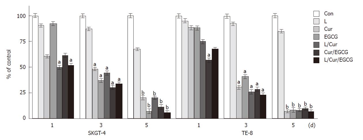
Figure 1 Suppression of esophageal cancer cell growth by curcumin, (-)-epigallocatechin-3-gallate, lovastatin and their combinations.
Esophageal cancer SKGT-4 and TE-8 cells were grown in monolayer overnight and then treated with or without curcumin (Cur) (40 μmol/L), (-)-epigallocatechin-3-gallate (EGCG) (40 μmol/L), lovastatin (L) (4 μmol/L) and their combinations for up to 5 d. Methyl thiazolyl tetrazolium assays were then carried out to detect changes in cell viability (see Methods section). The experiments were repeated three times and the results are summarized as a % of the control (Con) (mean ± SD) and analyzed statistically using the Student’s t test. aP < 0.05, bP < 0.01.
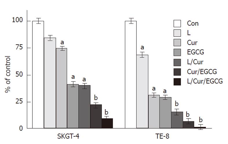
Figure 2 Suppression of tumor cell invasion by curcumin, (-)-epigallocatechin-3-gallate, lovastatin and their combinations.
Esophageal cancer SKGT-4 and TE-8 cells were grown and treated with or without curcumin (Cur) (40 μmol/L), (-)-epigallocatechin-3-gallate (EGCG) (40 μmol/L), lovastatin (L) (4 μmol/L) and their combinations in monolayer for 3 d and then subjected to cell invasion assays in Boyden chambers containing Matrigel for 48 h. The invasive cells were stained with 1% crystal violet solution, counted and the results are summarized as a % of the control (Con) (mean ± SD). The data were then analyzed statistically using the Student’s t test. aP < 0.05, bP < 0.01.
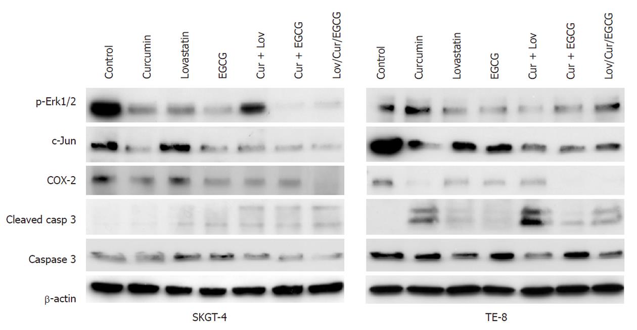
Figure 3 Modulation of gene expression by curcumin, (-)-epigallocatechin-3-gallate, lovastatin and their combinations.
Esophageal cancer SKGT-4 and TE-8 cells were grown in monolayer overnight and treated with or without curcumin (Cur) (40 μmol/L), (-)-epigallocatechin-3-gallate (EGCG) (40 μmol/L), lovastatin (Lov) (4 μmol/L) and their combinations for 2 d and total cellular protein was extracted from the cells and subjected to Western blotting analysis of gene expression. Erk1/2: Extracellular-signal-regulated kinases; COX-2: Cyclooxygenase-2.
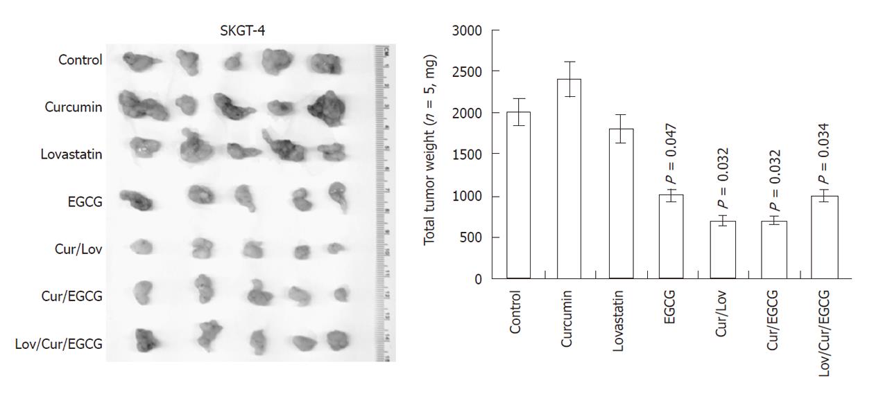
Figure 4 Inhibition of esophageal cancer cell growth by curcumin, (-)-epigallocatechin-3-gallate, lovastatin and their combinations in nude mouse xenografts.
Esophageal adenocarcinoma SKGT-4 cells were inoculated subcutaneously into nude mice (5 per group). Two days before tumor cell injection, the mice started treatment with or without curcumin (Cur) (50 μg/kg per day), (-)-epigallocatechin-3-gallate (EGCG) (50 μg/kg per day), lovastatin (Lov) (50 μg/kg per day) and their combinations (the same doses as given individually) for 30 d (5 d/wk by oral gavage). At the end of the experiments, the xenograft tumor mass was isolated and weighed and summarized.
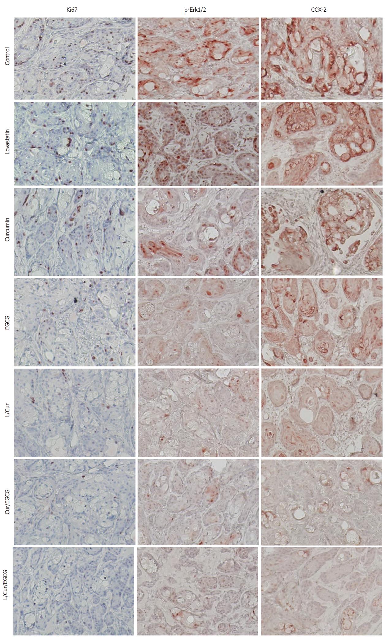
Figure 5 Reduced expression of Ki67, phosphorylated extracellular-signal-regulated kinases and cyclooxygenase-2 in xenografts following treatment of mice with or without curcumin, (-)-epigallocatechin-3-gallate, lovastatin and their combinations.
Tumor cell xenografts obtained from the nude mouse experiments were processed and subjected to immunohistochemical analyses of Ki67, phosphorylated extracellular-signal-regulated kinases (Erk1/2) and cyclooxygenase-2 (COX-2) expression. Representative images were obtained in each treatment group. EGCG: (-)-epigallocatechin-3-gallate; L: Lovastatin; Cur: Curcumin.
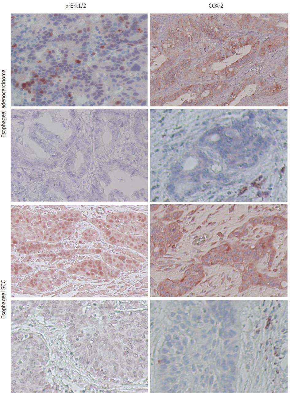
Figure 6 Expression of phosphorylated extracellular-signal-regulated kinases and cyclooxygenase-2 in esophageal cancer specimens.
Paraffin sections of esophageal cancer tissues were immunostained with anti-phosphorylated extracellular-signal-regulated kinases (Erk1/2) or cyclooxygenase-2 (COX-2) antibody. Representative images were obtained from these tissue sections. SCC: Squamous cell carcinoma.
- Citation: Ye F, Zhang GH, Guan BX, Xu XC. Suppression of esophageal cancer cell growth using curcumin, (-)-epigallocatechin-3-gallate and lovastatin. World J Gastroenterol 2012; 18(2): 126-135
- URL: https://www.wjgnet.com/1007-9327/full/v18/i2/126.htm
- DOI: https://dx.doi.org/10.3748/wjg.v18.i2.126


