Copyright
©2012 Baishideng Publishing Group Co.
World J Gastroenterol. Apr 28, 2012; 18(16): 1987-1990
Published online Apr 28, 2012. doi: 10.3748/wjg.v18.i16.1987
Published online Apr 28, 2012. doi: 10.3748/wjg.v18.i16.1987
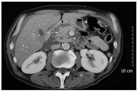
Figure 1 Preoperative abdominal computed tomography image showing inflammatory changes surrounding the head of the pancreas (arrow).
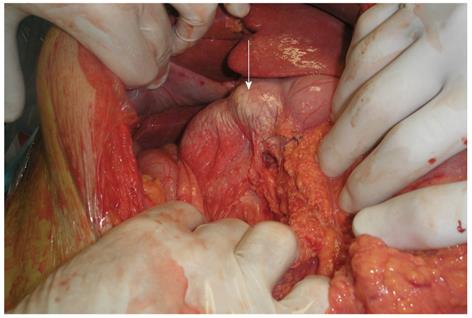
Figure 2 Intraoperative photo showing the milky-like appearance of the wall of the distal portion of the stomach and the duodenum due to congestion of the intestinal lymphatic drainage (arrow).
- Citation: Georgiou GK, Harissis H, Mitsis M, Batsis H, Fatouros M. Acute chylous peritonitis due to acute pancreatitis. World J Gastroenterol 2012; 18(16): 1987-1990
- URL: https://www.wjgnet.com/1007-9327/full/v18/i16/1987.htm
- DOI: https://dx.doi.org/10.3748/wjg.v18.i16.1987


