Copyright
©2011 Baishideng Publishing Group Co.
World J Gastroenterol. Feb 21, 2011; 17(7): 828-834
Published online Feb 21, 2011. doi: 10.3748/wjg.v17.i7.828
Published online Feb 21, 2011. doi: 10.3748/wjg.v17.i7.828
Figure 1 Computerized tomography.
A: Computerized tomography (CT) abdomen showing a patient with rectal cancer having liver metastasis and ascites; B: CT Chest showing a patient with rectal cancer having lung metastasis.
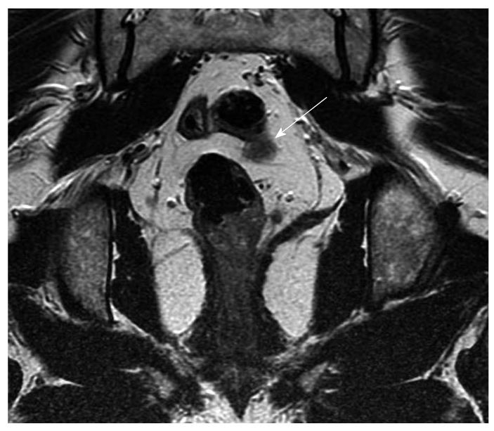
Figure 2 Coronal magnetic resonance imaging (arrow) showing possible lymph node or early vascular involvement.
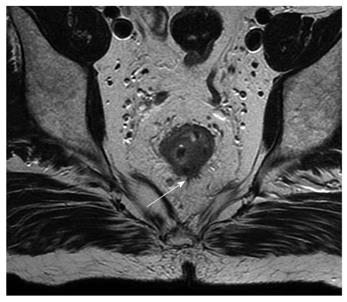
Figure 3 Magnetic resonance imaging (arrow) showing possible extension beyond the muscularis propria, radiologically staged as early T3.
Figure 4 Coronal T2 W magnetic resonance imaging (arrow) showing the intact muscularis propria in a patient with rectal cancer.
Radiologically staged as T1 or T2.
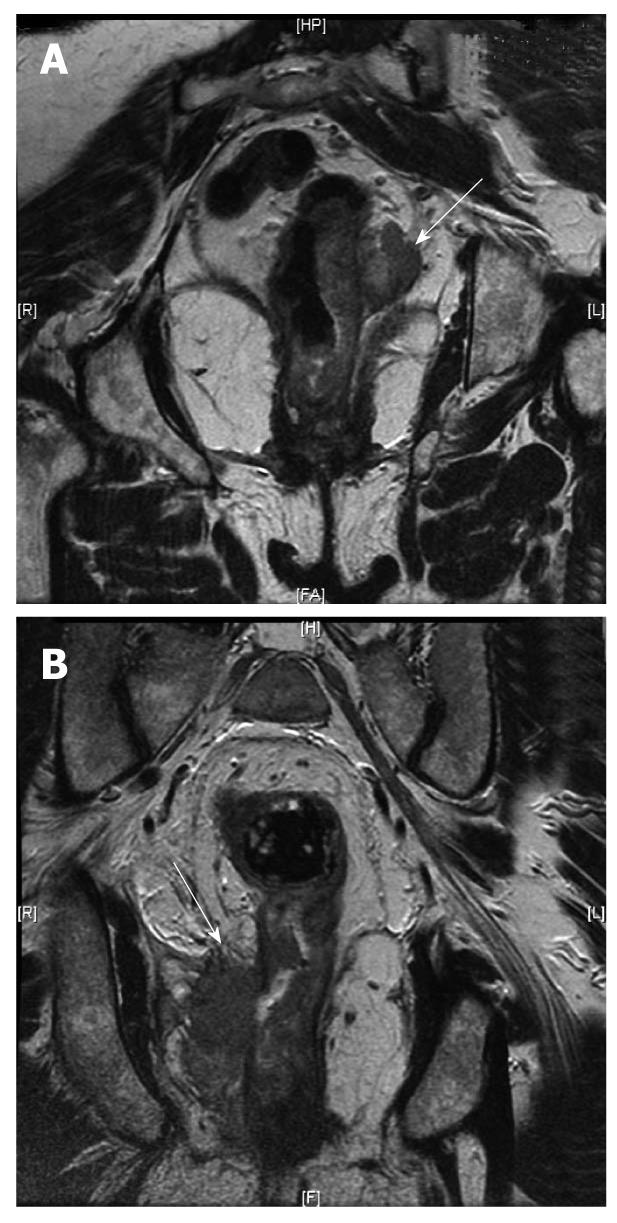
Figure 5 Magnetic resonance imaging.
A: Magnetic resonance imaging (MRI) (arrow) showing the rectal cancer involving the circumferential resection margin; B: MRI (arrow) showing the rectal cancer invading the ischiorectal fat on the right (T4).
- Citation: Samee A, Selvasekar CR. Current trends in staging rectal cancer. World J Gastroenterol 2011; 17(7): 828-834
- URL: https://www.wjgnet.com/1007-9327/full/v17/i7/828.htm
- DOI: https://dx.doi.org/10.3748/wjg.v17.i7.828


